









Surgeries y Uteruses
“Complex uterine malformation, reconstructive surgery and a successful pregnancy”
Extract:
A 33-year-old patient with a complex uterine malformation requiring a comprehensive approach to achieve a safe pregnancy. The case presents an added complication due to previous surgery, which has altered the anatomy and clinical presentation.
History:
A 33-year-old woman with primary infertility for one year. Regular periods, not heavy, not painful. Only one functional ovary. As a teenager, she had regular, painful and debilitating periods that led her to A&E on several occasions. She has been taking contraceptive pills for one year.
No previous treatments have been undertaken
Diagnosis:
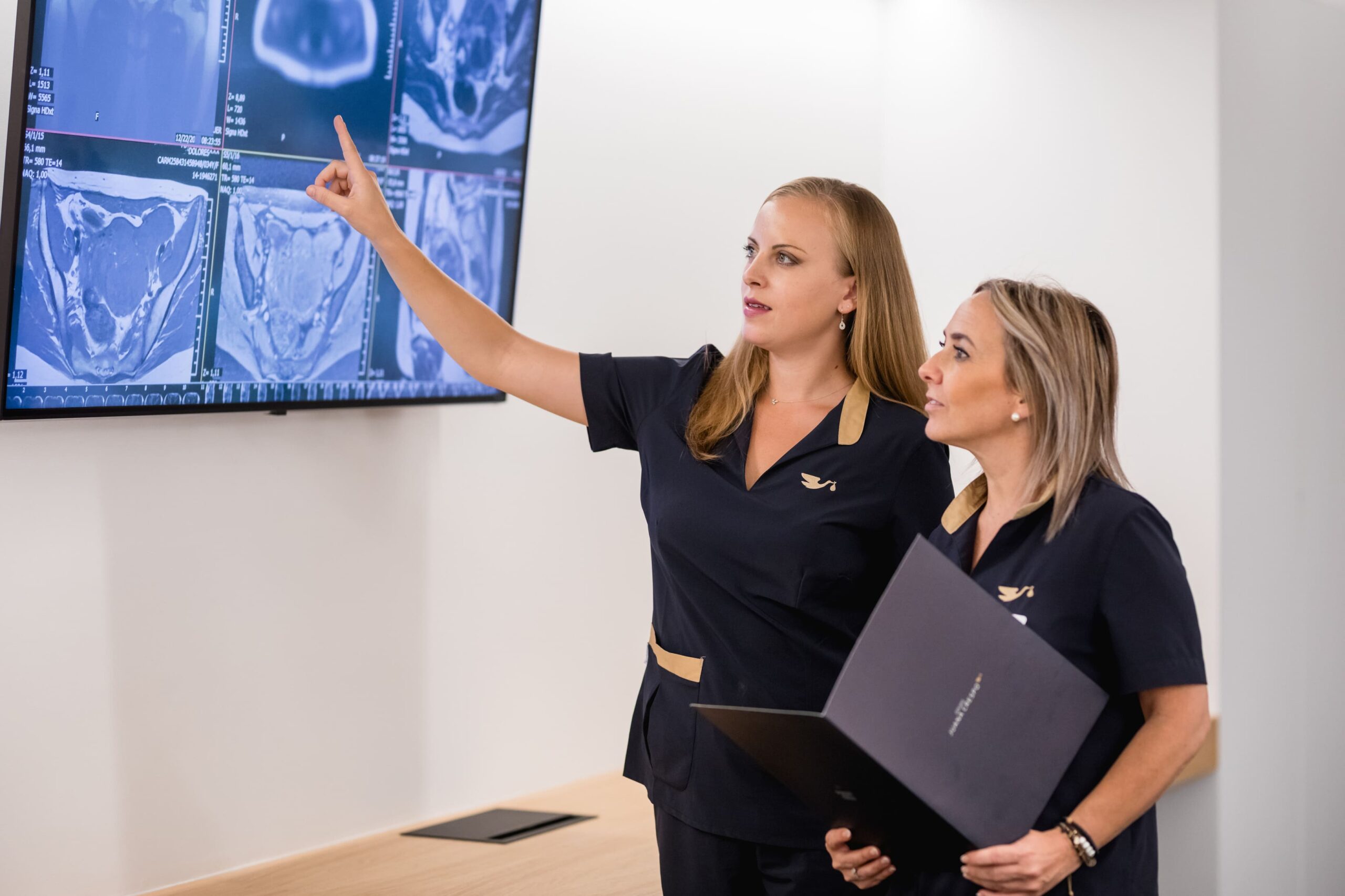
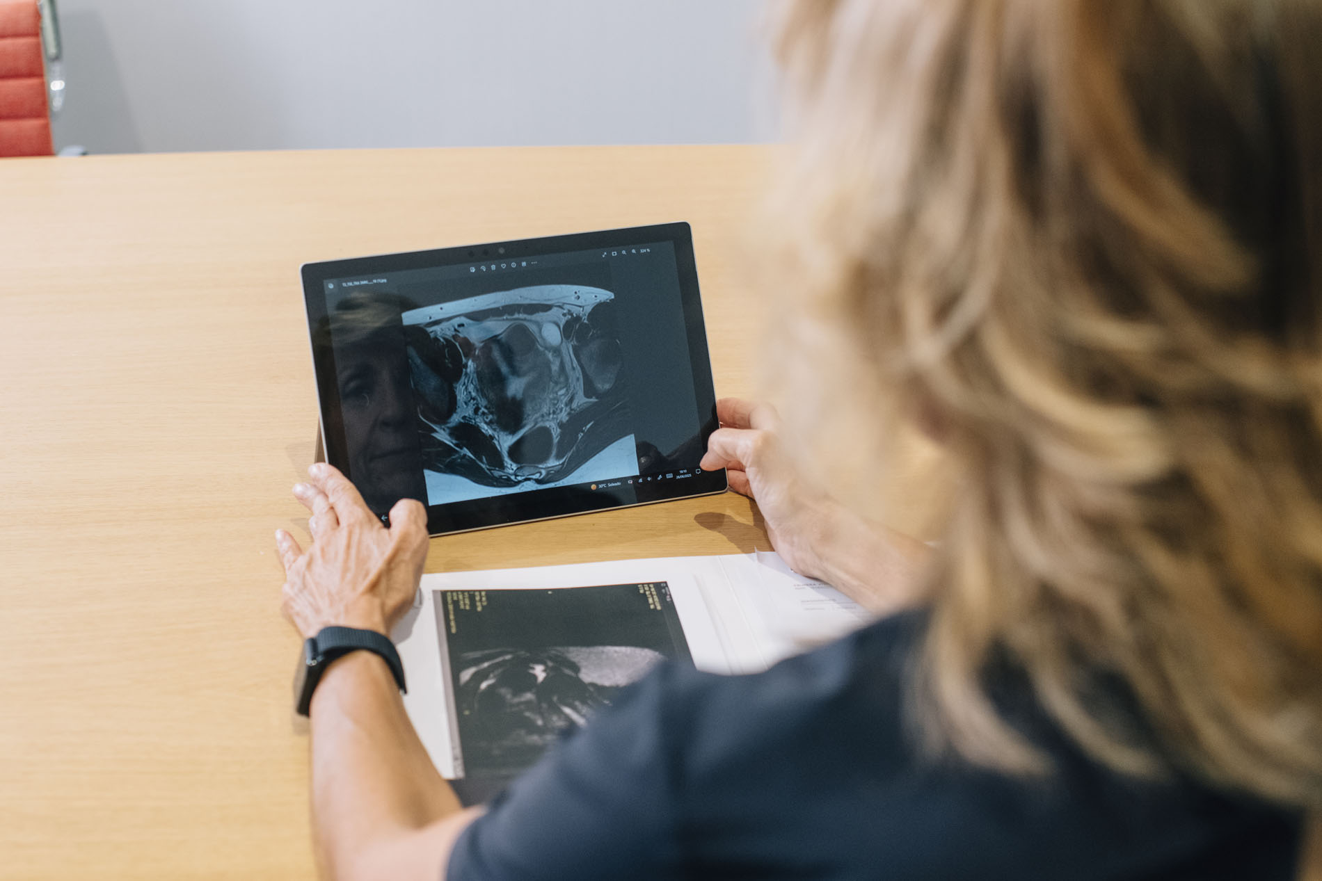
A new comprehensive assessment was carried out using
- High-resolution gynaecological ultrasound.
- Pelvic MRI.
- Diagnostic hysteroscopy.
All of this concludes that the patient presents with: a congenital malformation called unicornuate uterus, with a left uterine remnant that had no outlet to the cervix. Each month, menstrual blood became trapped in this closed cavity, causing:
- Intense pain.
- Progressive inflammation.
- Development of secondary adenomyosis.
Treatment plan:
A treatment plan is designed to meet two objectives:
- To definitively resolve the anatomical problem and the pain
- To preserve and optimise future reproductive capacity
For this reason, we structured the treatment into three phases.
Phase 1: Embryo preservation prior to surgery
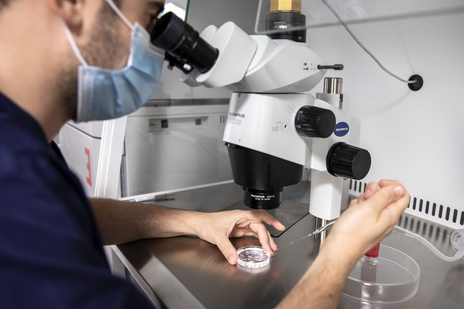
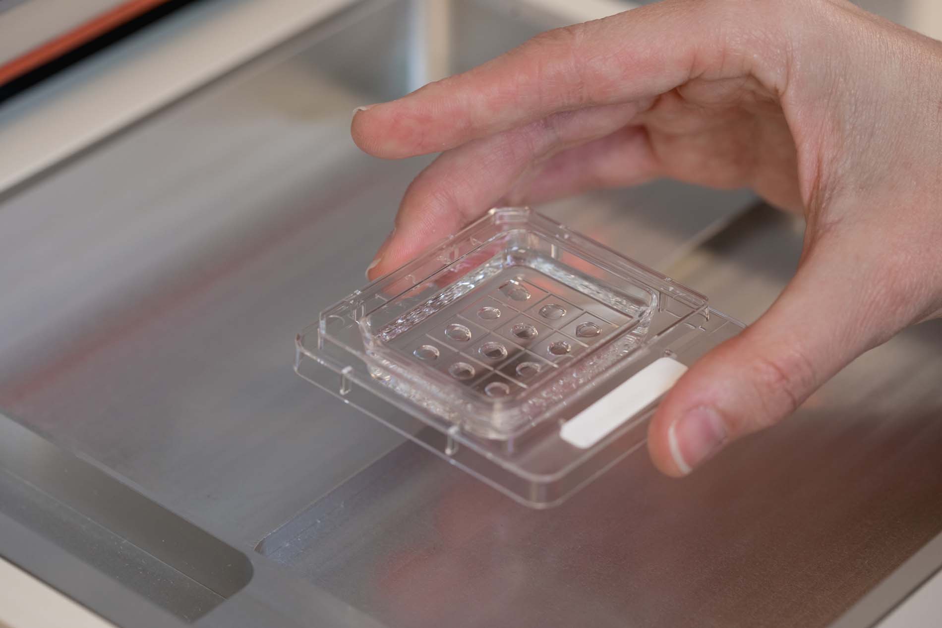
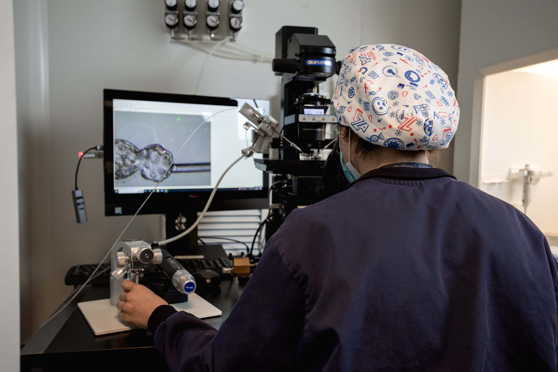
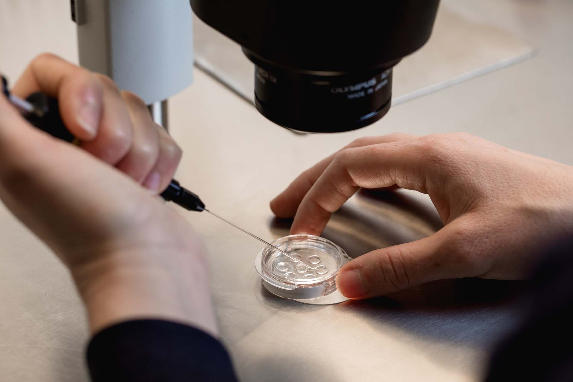
The strategy was based on achieving a good embryo yield; 13 oocytes were retrieved, resulting in 3 chromosomally normal embryos analysed by PGTA and 1 low-grade mosaic.
This allowed us to have healthy embryos before performing surgery on the uterus, increasing safety and reducing subsequent risks.
Phase 2: Highly complex reconstructive surgery
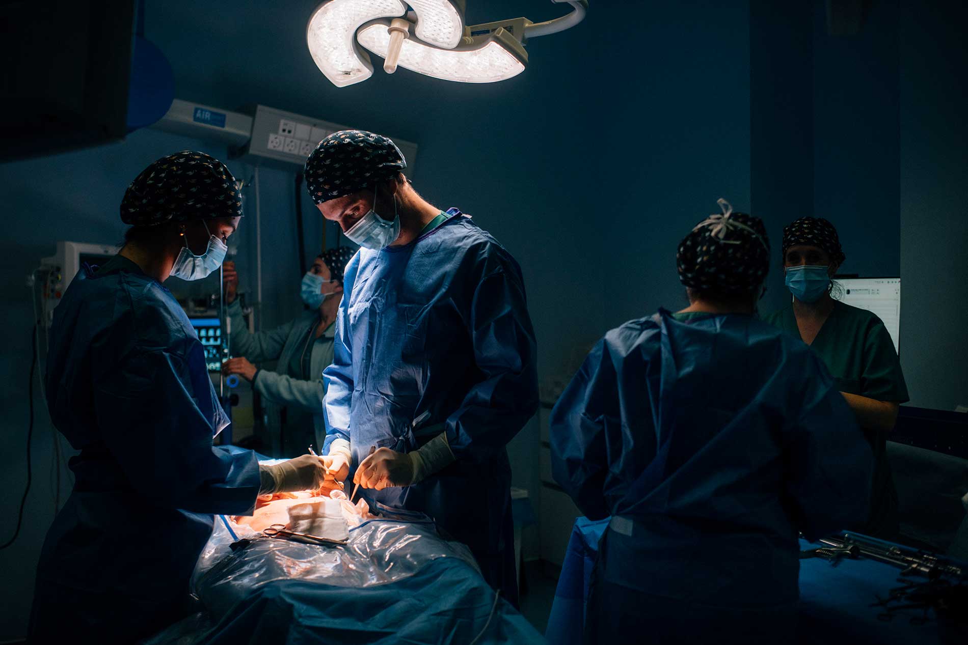
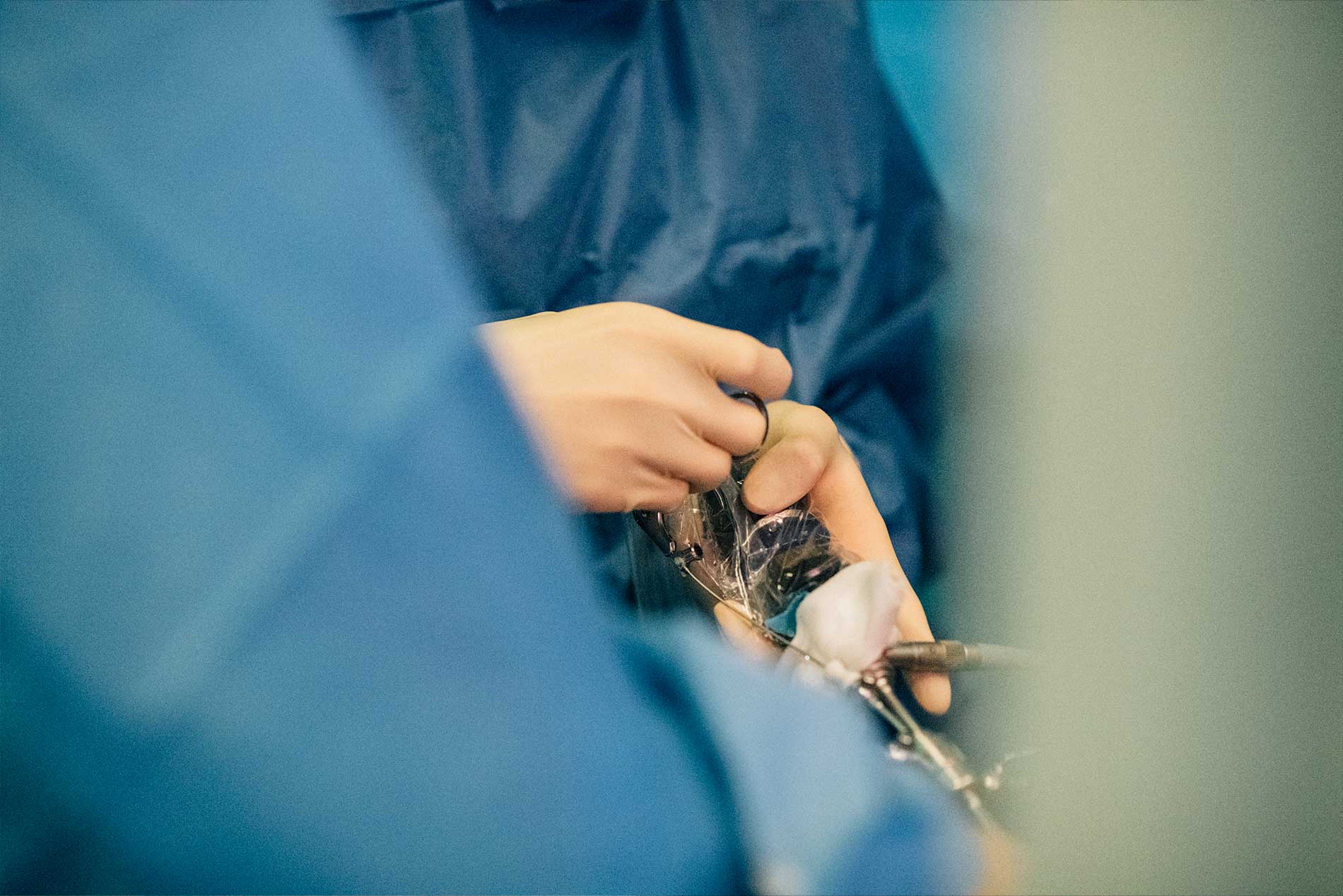
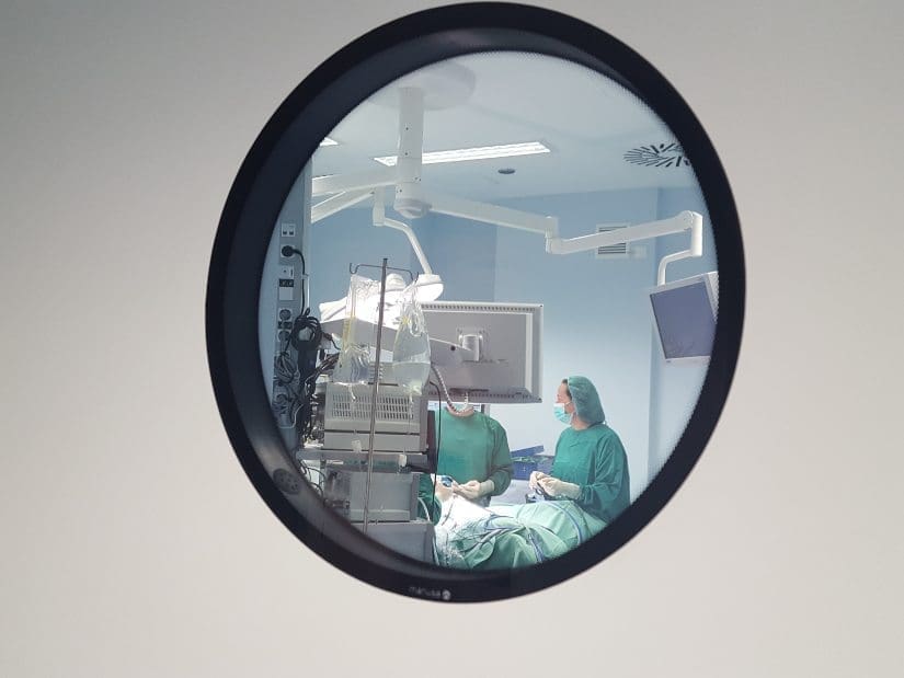
Advanced laparoscopic surgery was performed.
- Complete resection of the left rudimentary horn (left hemihysterectomy).
- Right salpingectomy.
- Left ovarian fixation.
Why was this surgery necessary?
- To remove the cavity where blood was accumulating.
- To halt the progression of adenomyosis.
- To leave a uterus that is anatomically safe for pregnancy.
Phase 3: Hysteroscopic optimisation
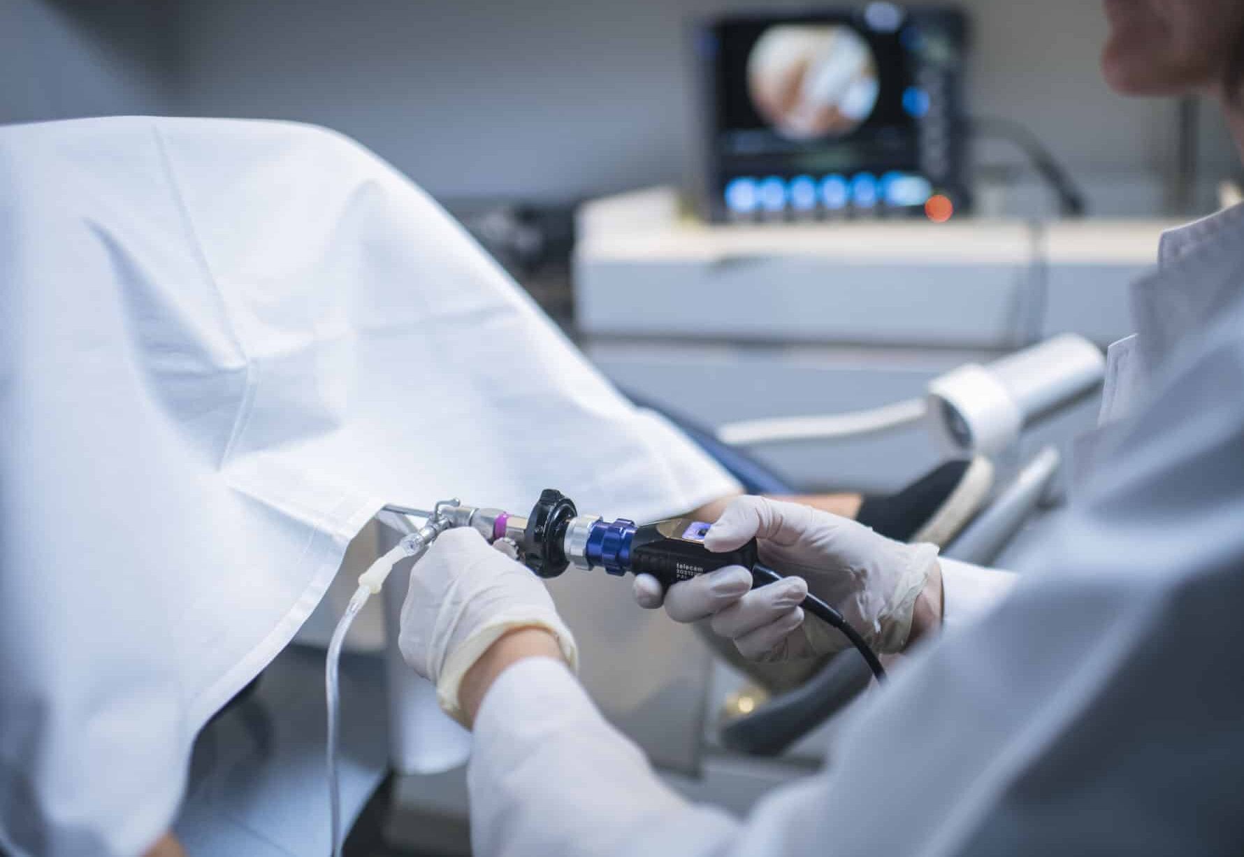
Months later, we performed a surgical hysteroscopy, a guided uterine repair microsurgery to:
- Slightly enlarge the cavity of the unicornuate uterus to improve uterine distensibility and functionality
This step is essential in complex cases: it is not enough to remove the malformation; it is necessary to prepare the uterus to receive an embryo.
Phase 4: Endometrial preparation with embryo transfer
Following six months of surgical recovery and highly personalised endometrial preparation:
- We transferred a single chromosomally normal embryo.
- Implantation was achieved.
- The pregnancy progressed normally.
- Discharge from obstetric care.
Conclusion
At Equipo Juana Crespo, we treat complex infertility from a holistic perspective:
- Advanced anatomical diagnosis.
- Specialised reconstructive gynaecological surgery.
- Transfer at the optimal time.
This case demonstrates that, even in the face of severe uterine malformation with secondary adenomyosis, it is possible to achieve a successful pregnancy when treatment is correctly sequenced and personalised.


