





Adenomyosis
“When pregnancy does not reach full term following multiple pregnancy losses and cervical insufficiency: a comprehensive strategy for achieving a live birth”
A complex reproductive history
A 40-year-old woman with a history of infertility and recurrent pregnancy loss since the age of 23.
Throughout her reproductive history, she had experienced 15 pregnancies: 13 spontaneous and 2 following assisted reproductive technology. Of the spontaneous pregnancies, 7 resulted in preterm births or late miscarriages, between 16 and 23 weeks’ gestation, due to cervical incompetence.
Following several treatment attempts, including three cervical cerclages and one abdominal cerclage, as well as assisted reproduction treatments, the patient presented for a further comprehensive assessment of her case.
Obstetric history
The pregnancy losses occurred recurrently in the second trimester: Furthermore, she experienced two biochemical pregnancies following frozen embryo transfers.
- Week 22, aged 23.
- Week 20, aged 25.
- Week 16, aged 26.
- Week 19, aged 27.
- Week 16, aged 30.
- Week 21, aged 31.
- Week 23, subsequently.
Surgical history and previous treatments
The patient had previously undergone three cervical cerclages, all of which were followed by further pregnancy losses, and an abdominal cerclage performed in 2019.
She had also required three dilation and curettage procedures.
Subsequently, after attempting to conceive again in 2022, she was unable to become pregnant spontaneously.
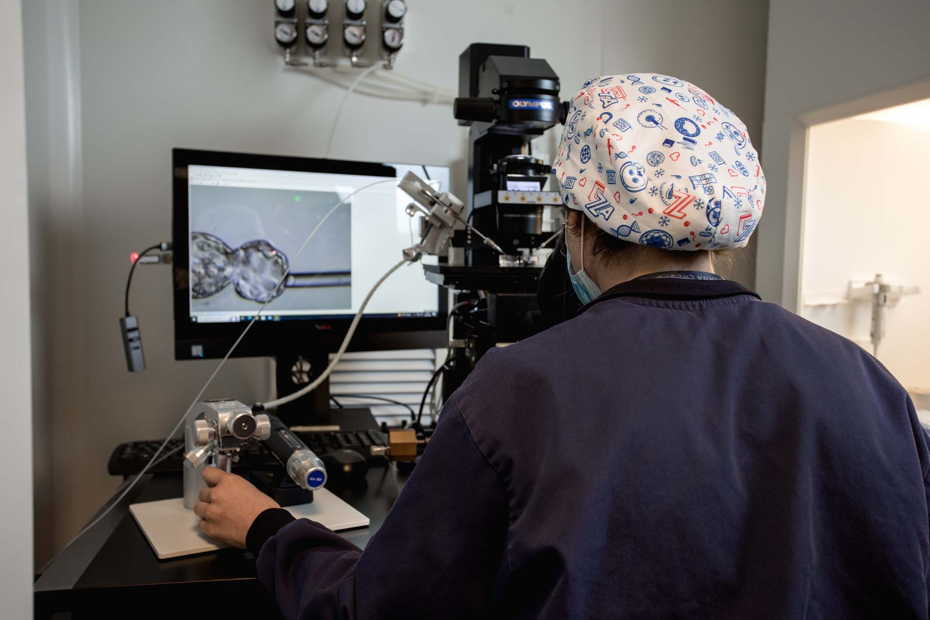
As part of assisted reproduction treatment, ICSI with PGT-A was performed, yielding two euploid embryos which were transferred on two occasions, both of which resulted in biochemical pregnancies.
Gynaecological examination
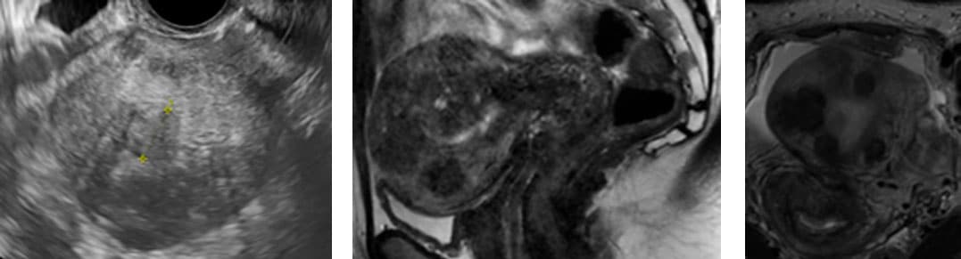
Ultrasound assessment revealed a polymyomatous uterus, with several fibroids located mainly in the anterior wall, some of which protruded into the cavity and disrupted the regularity of the endometrium.
Furthermore, significant adenomyosis was observed, particularly in the posterior wall, with a globular and pathological uterine morphology.
Another key finding was the persistence of cervical incompetence, with a previous cerclage situated in a low position.
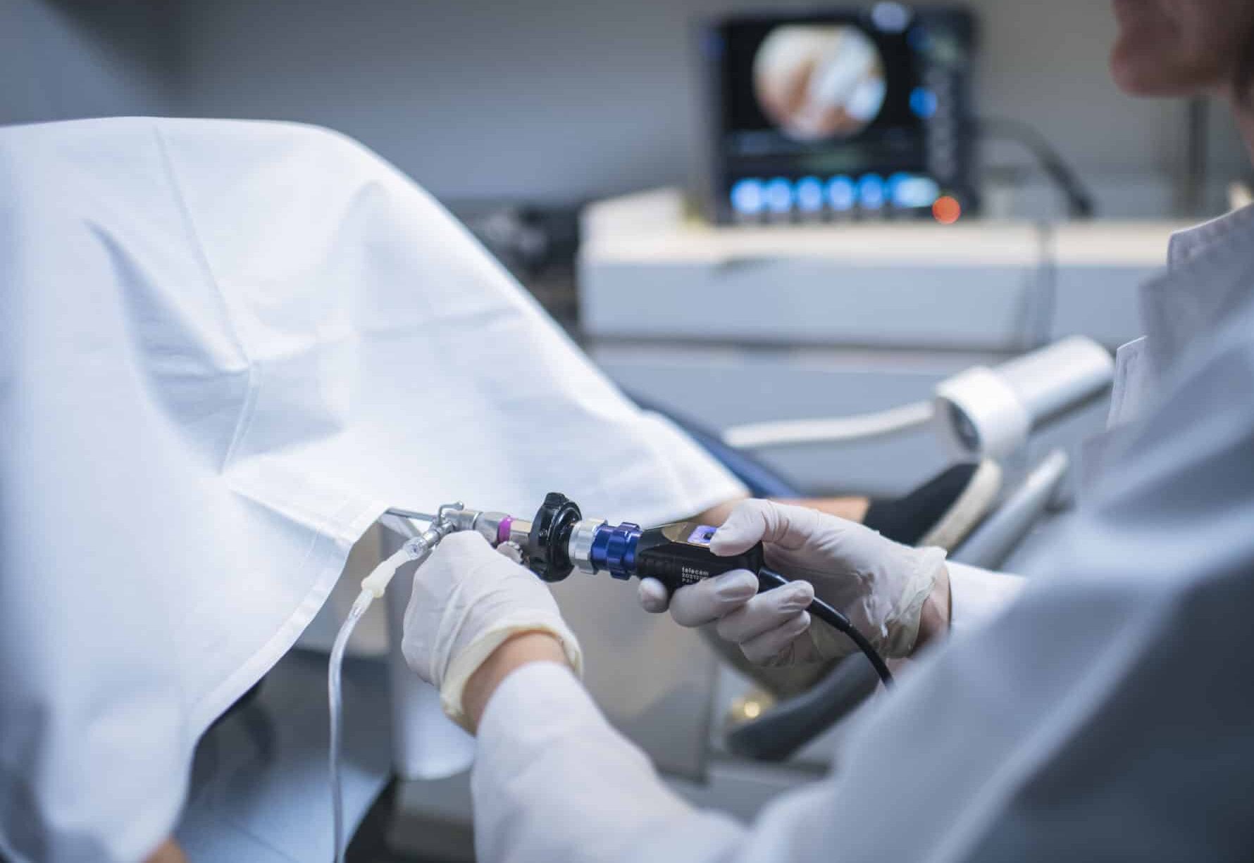
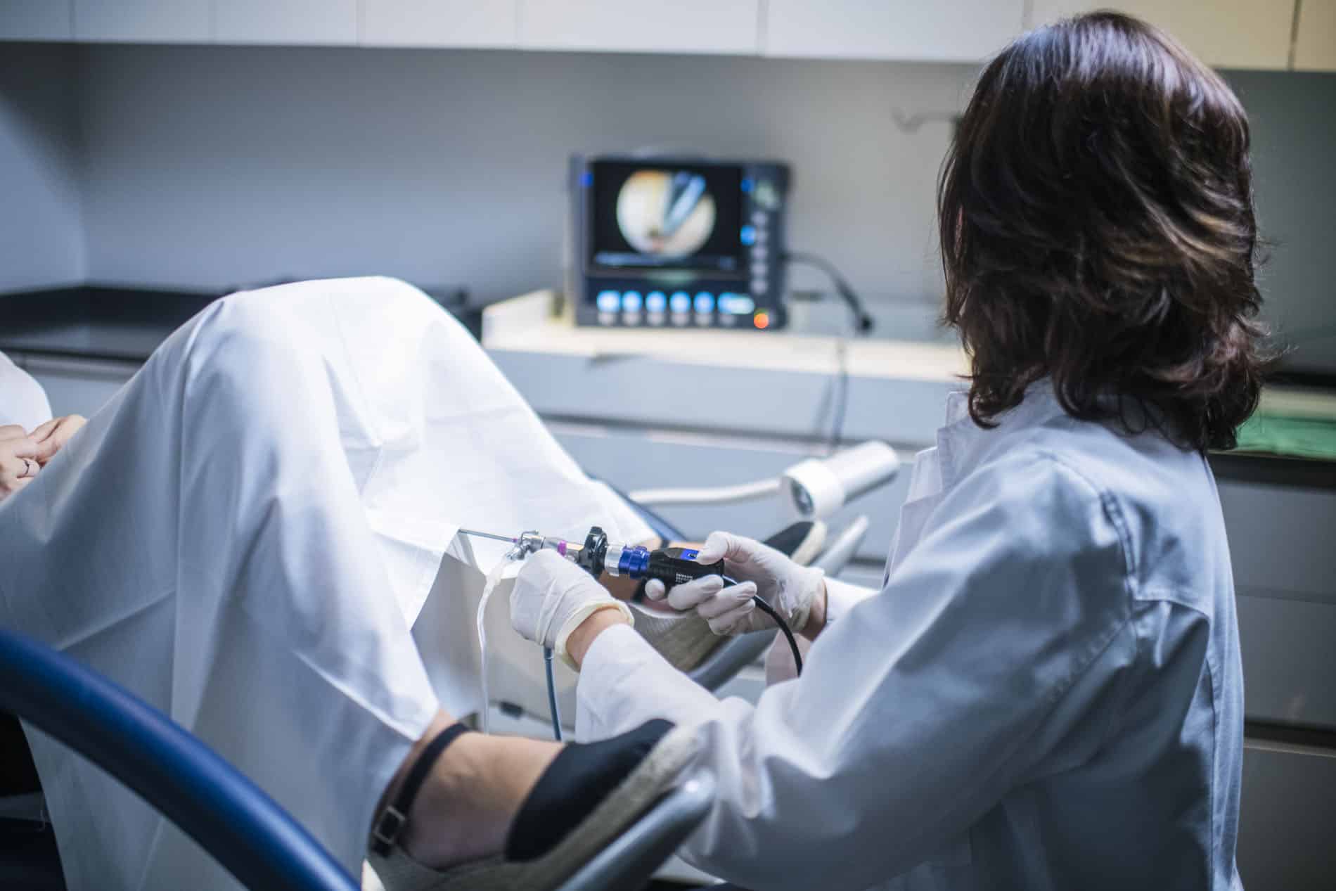
Diagnostic hysteroscopy confirmed the marked cervical incompetence. The inability to adequately distend the uterine cavity also allowed observation of the protrusion of one of the anterior wall fibroids into the cavity.
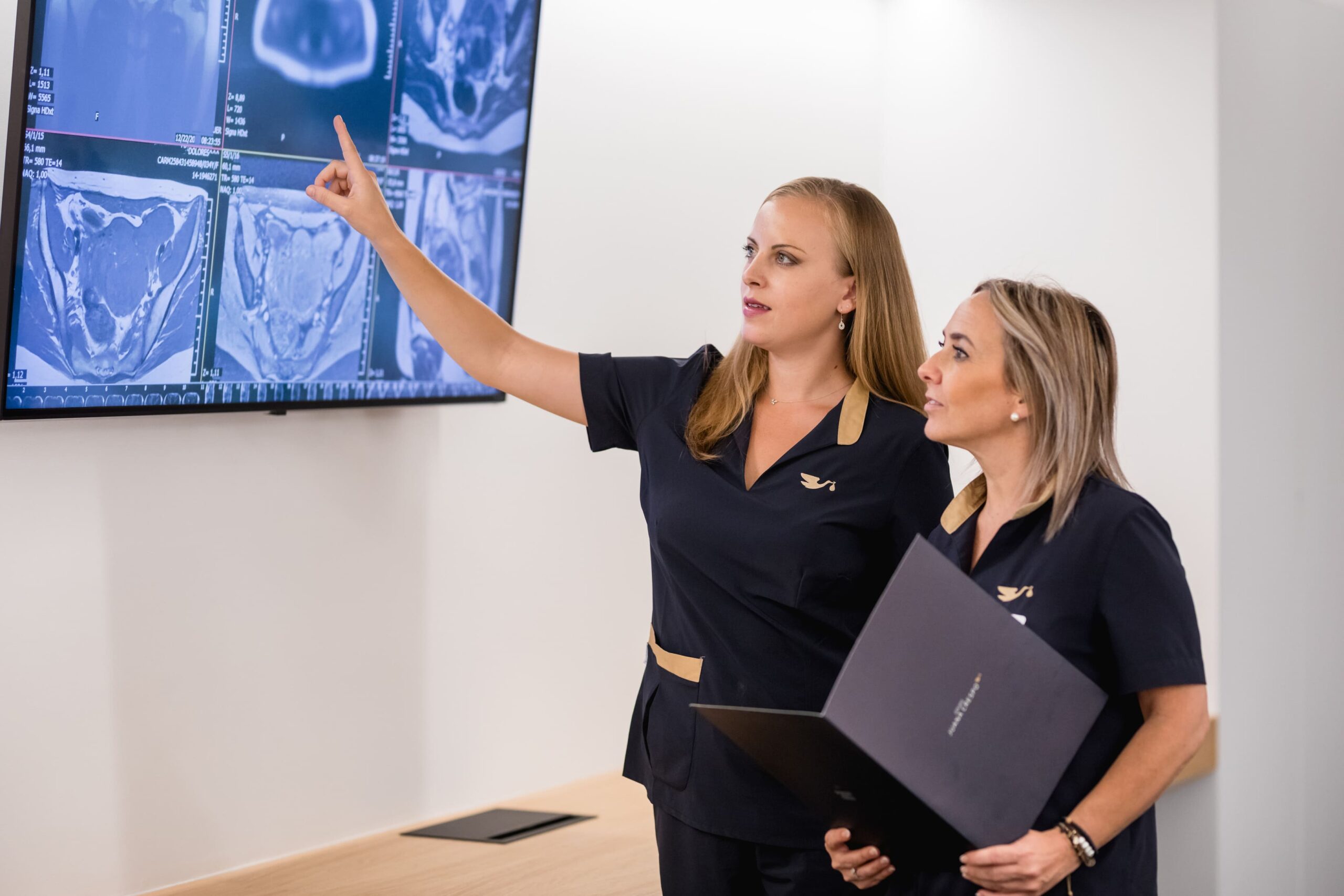
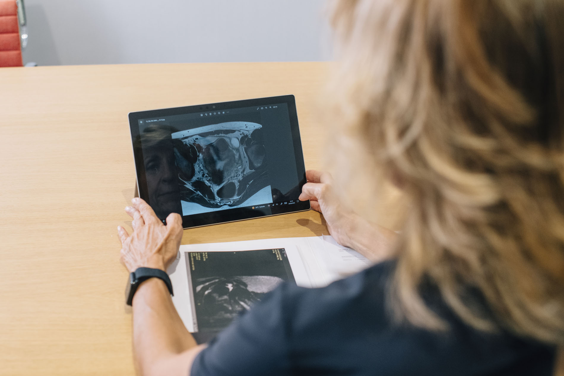
Magnetic resonance imaging confirmed the presence of a polyfibroids and adenomyotic uterus, with fibroids compromising and deforming the endometrial lining.
A personalised treatment strategy
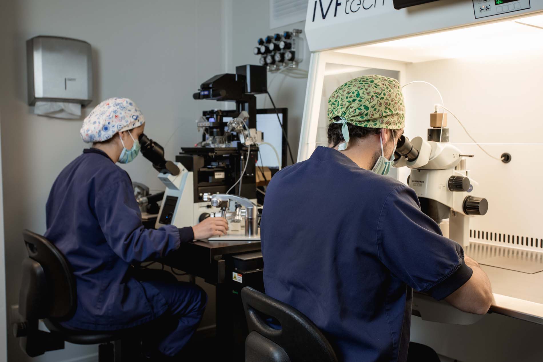
Given the complexity of the case, a sequential treatment strategy was devised, with the aim of optimising both the uterine anatomy and the conditions for a future pregnancy. Using ICSI and PGT-A, the following were obtained: In the first surgical stage, a laparoscopy was performed.
- Embryo selection
- 1 euploid embryo, D5 stage.
- 1 low-grade mosaic embryo.
- Laparoscopy, myomectomy and new abdominal cerclage
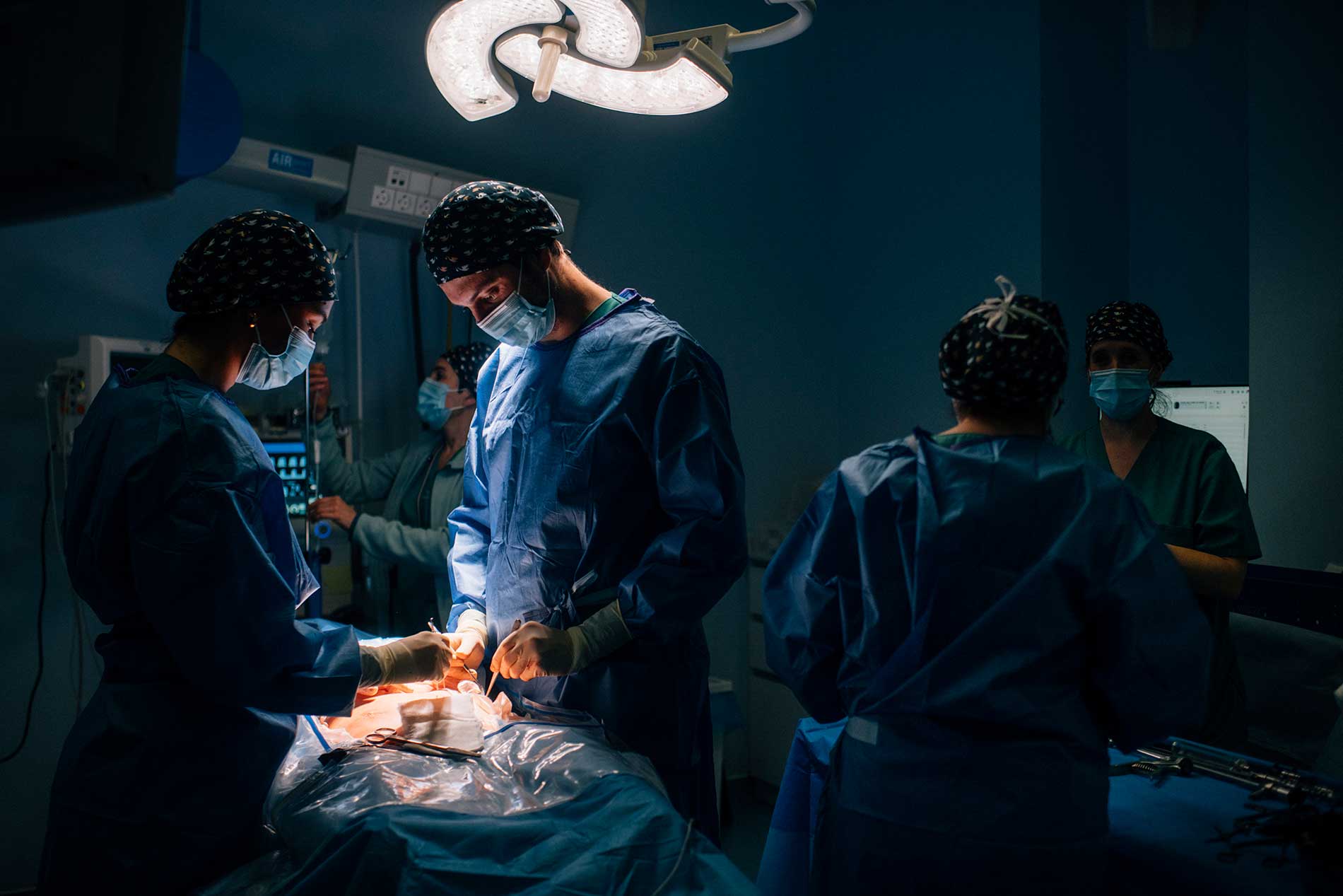
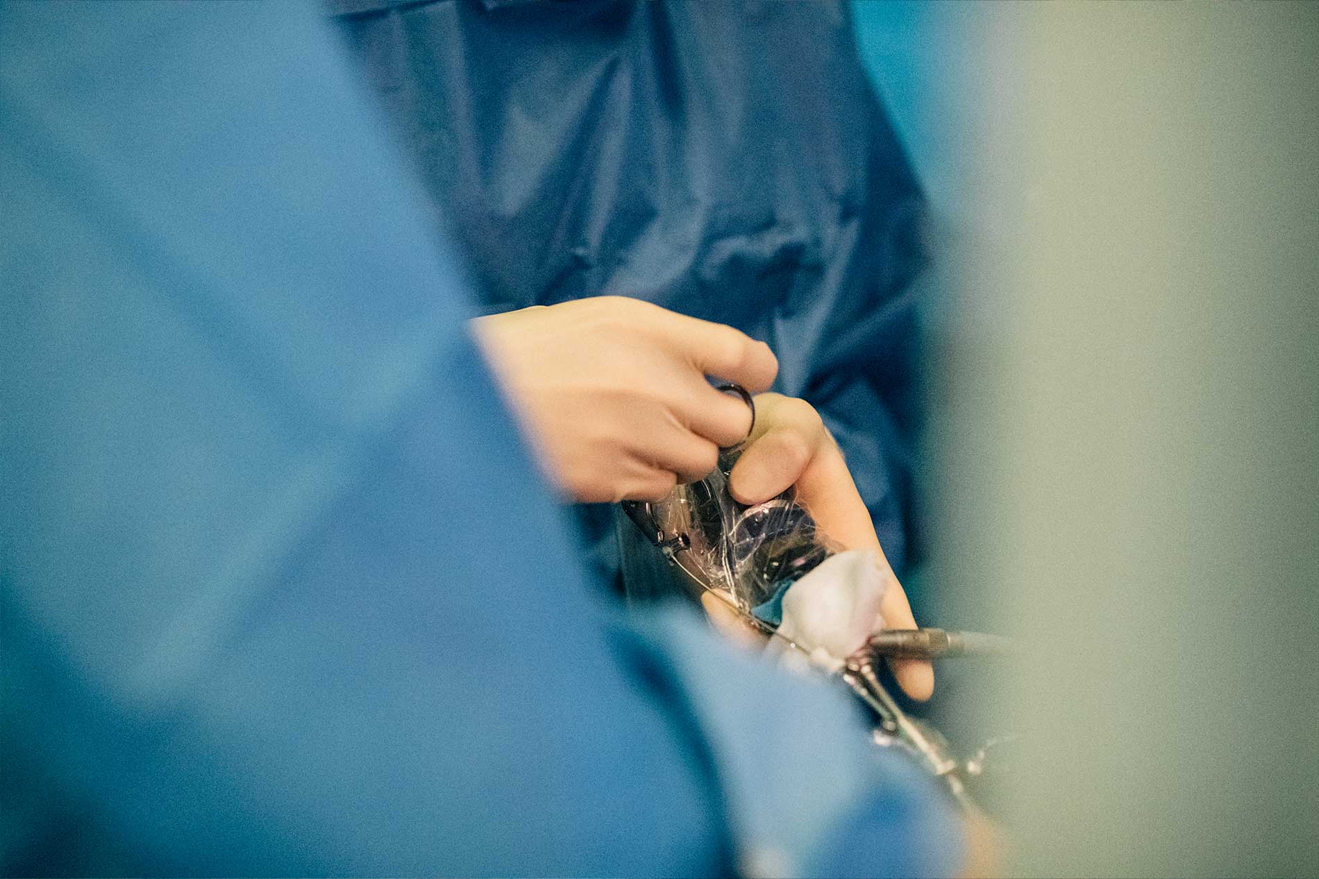
A polymyomatous and adenomyotic uterus was observed, with significant adenomyosis in the posterior wall and multiple fibroids predominantly in the anterior wall. The ovaries and fallopian tubes appeared normal.
During the procedure, the previous abdominal cerclage, performed with Mersilene, was identified in a low position.
A multiple myomectomy involving seven fibroids was performed, avoiding opening the uterine cavity, and the previous cerclage was removed.
Subsequently, a new abdominal cerclage was performed using Mersilene, positioned higher than the previous one, approximately 15–20 mm above it, with its correct position verified by ultrasound.
Following the surgery, anti-oestrogen treatment was initiated for three months, with the aim of promoting recovery following the myomectomy and treating the adenomyosis. Once the recovery period was complete, a second-stage surgical hysteroscopy was performed.
- Surgical hysteroscopy and treatment of adenomyosis
Intracavitary myomas were resected using hysteroscopic micro-scissors, followed by treatment of the adenomyosis lesion and the application of platelet-rich plasma (PRP).
Subsequently, medical treatment aimed at reducing the extent of adenomyosis affecting the posterior wall was continued, and the uterus was prepared for embryo transfer in a natural cycle.
Result:
Following completion of the therapeutic strategy and restoration of normal uterine anatomy, the first transfer of a euploid embryo was carried out.
The patient achieved a progressing pregnancy and subsequently a successful pregnancy.
The placenta was located in a high anterior position and, given the obstetric complexity and history of cervical incompetence, a caesarean section was scheduled for week 37 of gestation.
Conclusion
This case highlights the importance of approaching cases of high reproductive complexity from a comprehensive and personalised perspective, particularly when factors such as cervical incompetence, uterine myomatosis, adenomyosis and a prolonged history of pregnancy loss are present.
The complexity of this patient’s case required much more than a single treatment. It was necessary to meticulously analyse her uterine and cervical anatomy, identify the causes that might be interfering with the achievement and progression of the pregnancy, and design a surgical and reproductive strategy tailored to her needs.
The role of a surgical team specialising in complex gynaecological surgery and reproductive medicine was crucial in addressing each of these factors in a coordinated manner. Expertise in laparoscopic and hysteroscopic surgery enabled the team to perform a multiple myomectomy whilst preserving the uterine cavity, to modify and reposition the abdominal cerclage, and subsequently to treat the intracavitary pathology and adenomyosis.
Each decision was taken sequentially, assessing the outcome of each procedure before moving on to the next step. This planning made it possible to optimise the uterine anatomy, improve conditions for implantation and provide the embryo with the best possible conditions for a successful pregnancy.
The success of this case highlights that, when faced with a complex reproductive history, experience, surgical precision and the coordinated work of a specialised team can make all the difference.
The ultimate aim was not merely to achieve a pregnancy, but to transform a uterus with multiple risk factors into the most favourable environment possible for a successful birth.


