


Surgeries
“Fertility preservation in a patient with a giant myomatous uterus”
Abstract
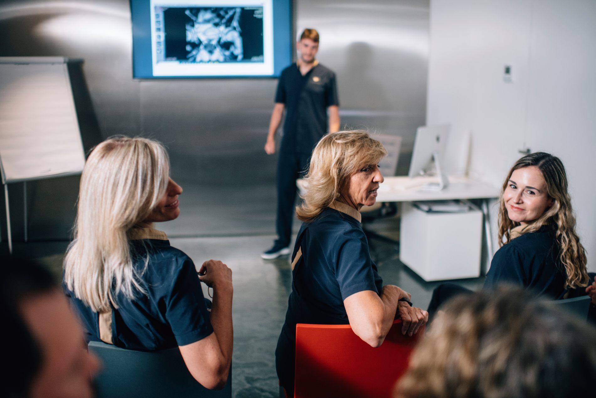
The Juana Crespo Team, a specialist assisted reproduction clinic, presents the case of a 38-year-old patient with a giant myomatous uterus who had been advised that a hysterectomy was her only option, as the multiple fibroids had completely distorted the endometrial cavity and made pregnancy impossible.
The multidisciplinary team of specialists designed a conservative approach that included IVF (in vitro fertilisation) with embryo vitrification, laparotomy myomectomy, hysteroscopic myomectomy and subsequent endometrial preparation. Ultimately, a successful pregnancy was achieved. This case demonstrates the viability of conservative reproductive strategies in anatomically complex situations.
Introduction
Uterine fibroids are the most common benign neoplasm of the female reproductive tract and can compromise fertility depending on their number, size and location. In cases of giant myomatous uteri, many patients are offered hysterectomy as the sole therapeutic option, which precludes any future reproductive options.
We present the case of a 38-year-old patient who attended our assisted reproduction clinic seeking conservative alternatives with the aim of preserving her reproductive potential.
Background
- 38-year-old patient.
- Desire for pregnancy
- At other centres, she had only been offered a hysterectomy due to the size of the fibroids and the surgical complexity.
Treatment Plan
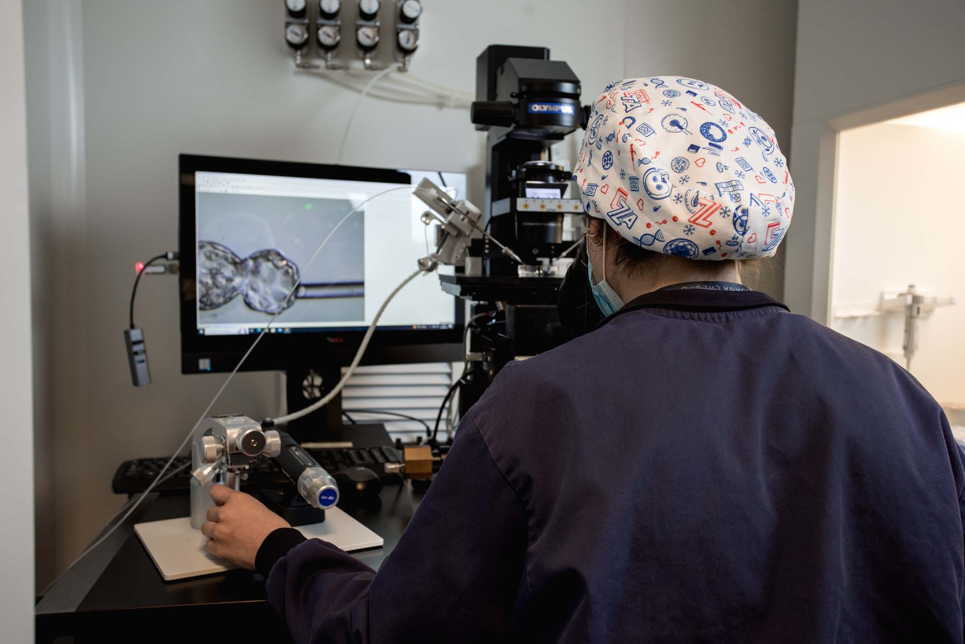
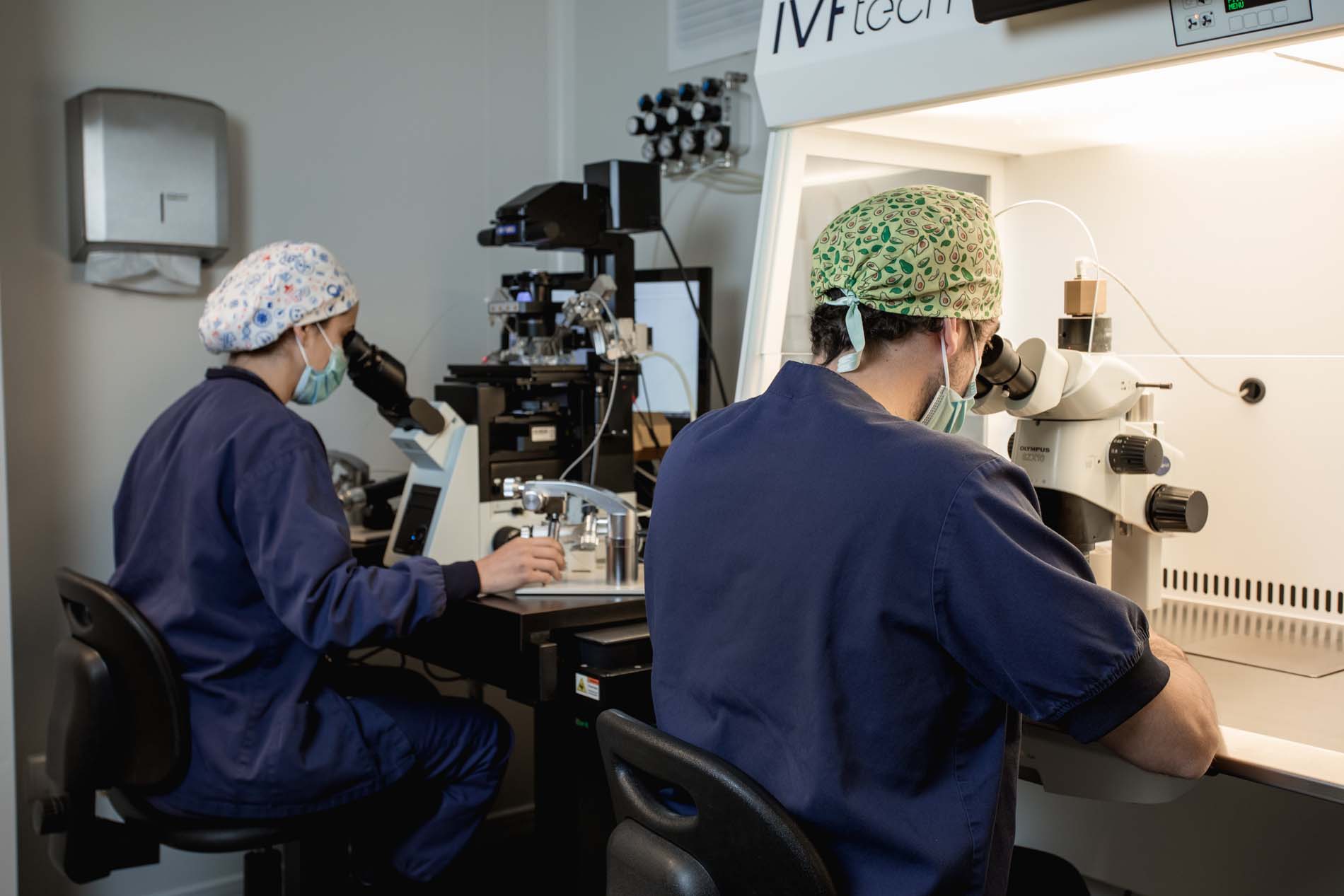
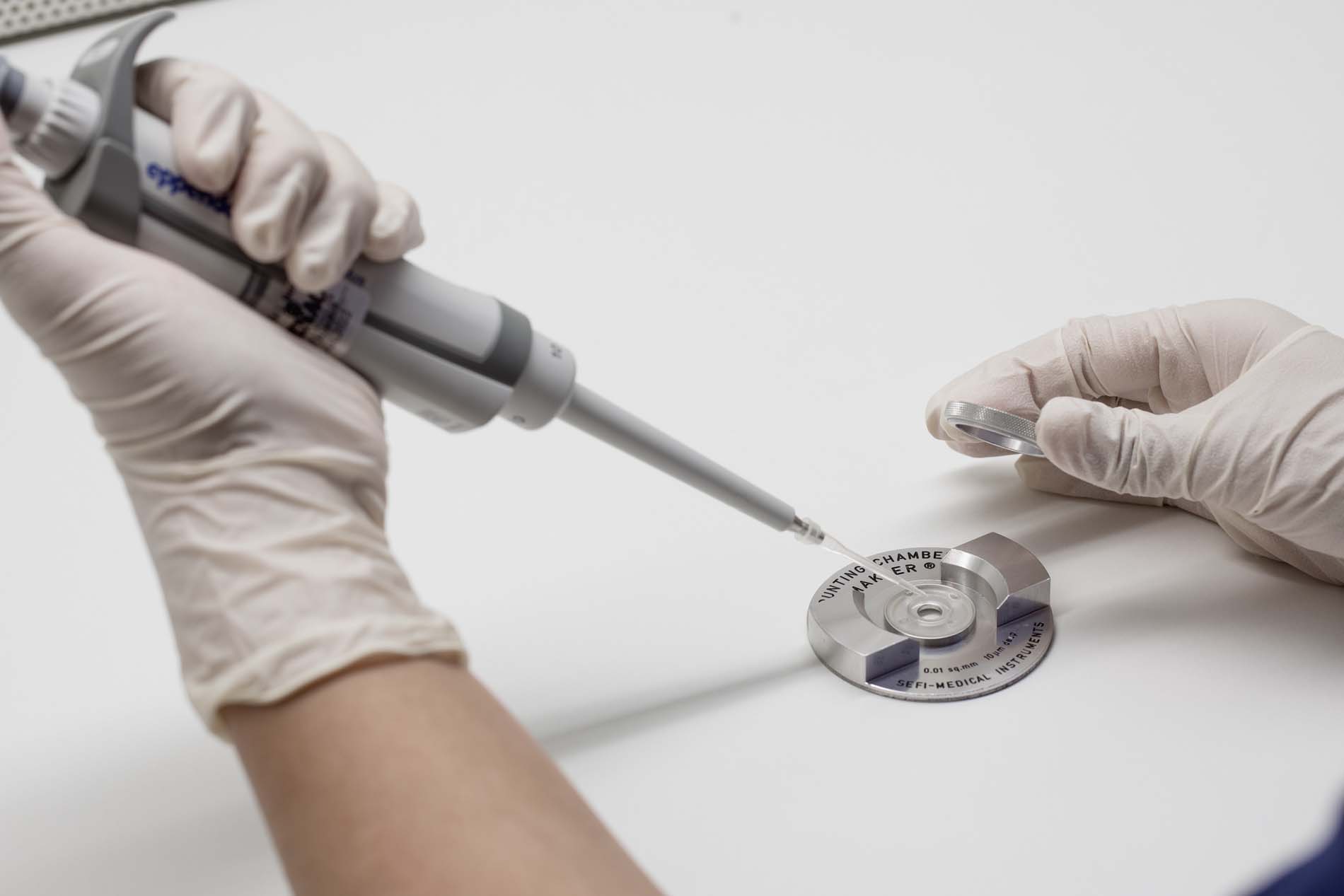
First step: preserving fertility through IVF and embryo vitrification
Before subjecting the uterus to reconstructive surgery, we carried out:
- Ovarian stimulation
- In vitro fertilisation
- Embryo vitrification to ensure future reproductive potential
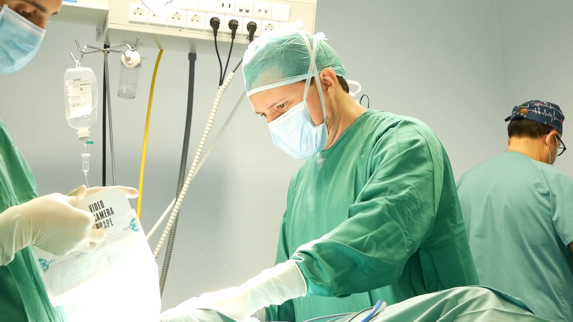
Laparotomy myomectomy: restoring the anatomy
The size of the uterus necessitated an open surgical approach. Upon opening the abdominal cavity, a massive cluster of fibroids was observed, responsible for the abdominal deformity visible preoperatively.
The surgery allowed for the removal of multiple myomatous masses and the restoration of the uterus’s general shape whilst preserving its functionality.
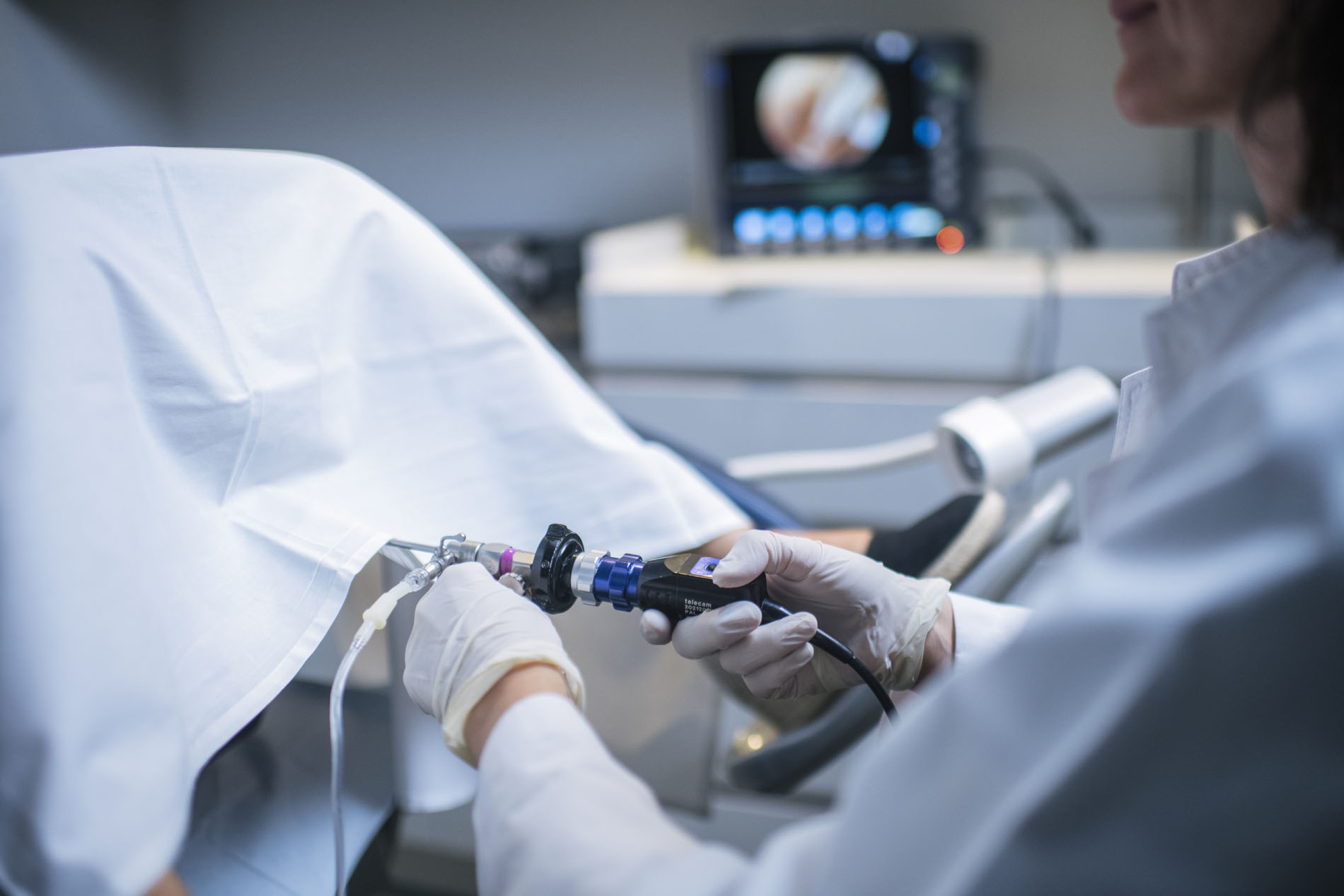
Second step: hysteroscopic myomectomy
Following recovery, we performed a hysteroscopic procedure to remove residual submucosal tissue and restore a functional endometrial cavity.
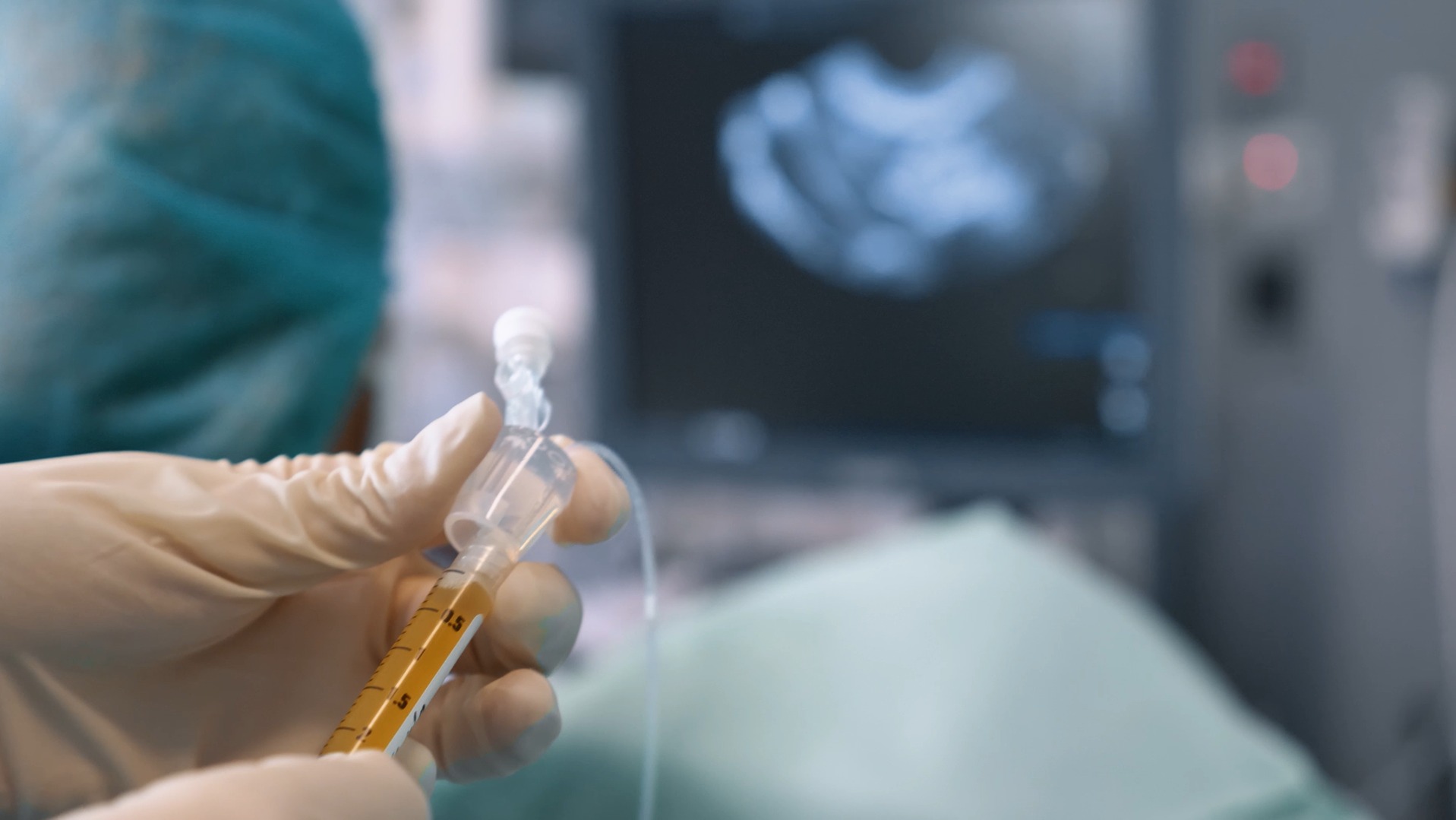
Final outcome: a successful pregnancy
After properly preparing the endometrium, one of the vitrified embryos was transferred. The result was a progressive pregnancy, confirming the success of the conservative approach.
This case demonstrates that, even in the presence of a giant myomatous uterus, it is possible to avoid a hysterectomy and preserve reproductive capacity when working with a team specialising in reproduction and advanced gynaecological surgery.
Innovation in the therapeutic approach:
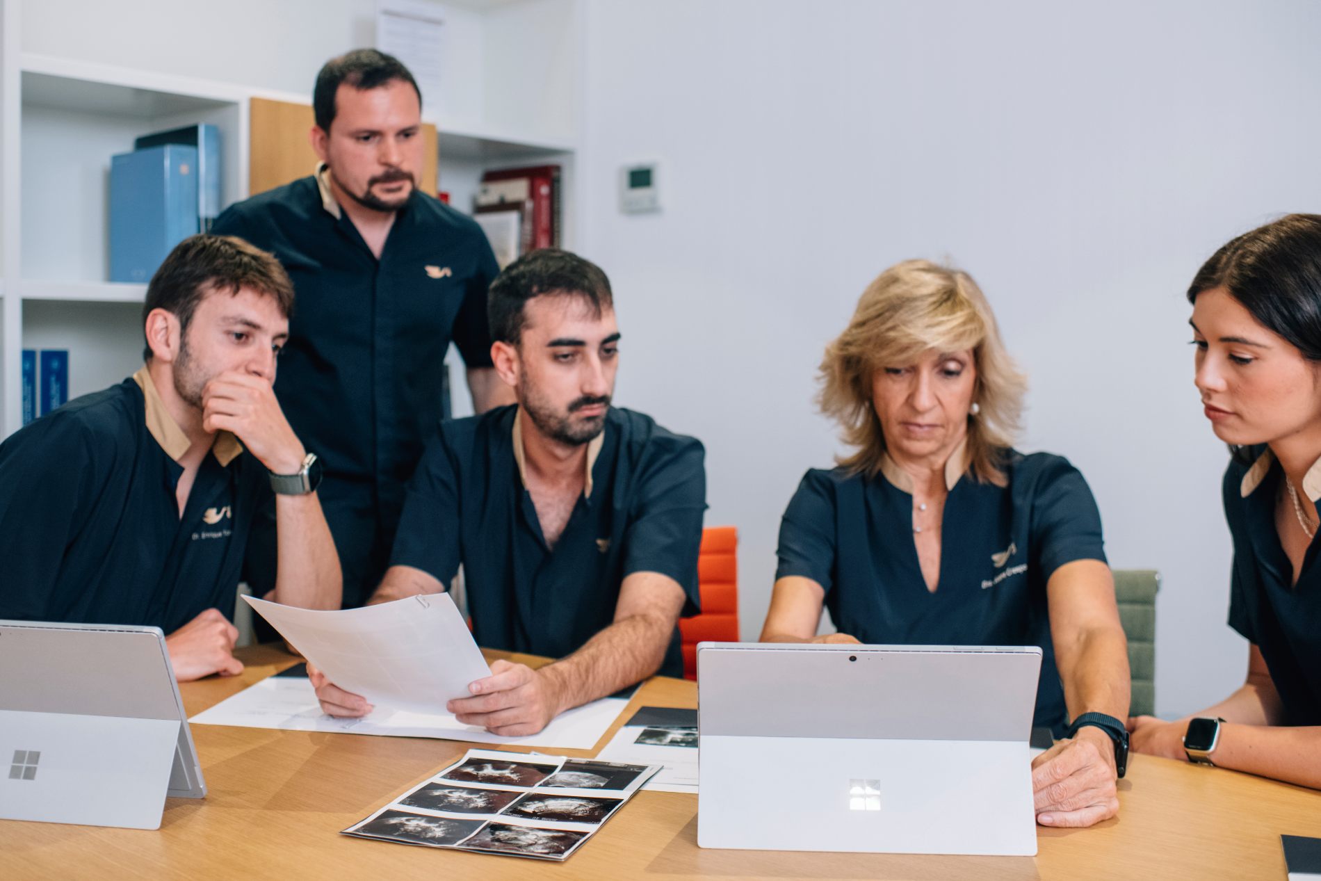
This case highlights:
- The importance of a multidisciplinary approach combining surgery and assisted reproduction.
- That even in extreme cases of a giant myomatous uterus, uterine preservation is possible.
- The usefulness of embryo vitrification in protecting fertility during complex surgical procedures.
- That sequential myomectomy (laparoscopic and hysteroscopic) can restore uterine function.
Conclusions
The combination of assisted reproduction techniques and conservative surgery made it possible to avoid a hysterectomy and achieve a pregnancy in a patient with a giant myomatous uterus. It is essential to offer conservative alternatives in specialist centres.