













Complex Punctures
“Fertility preservation: ovary-sparing surgery for an 11 cm tumour”

Abstract
A 30-year-old woman with primary infertility lasting one year and a desire to preserve her fertility. She had a single ovary following an oophorectomy at the age of 18 and, at the age of 28, a complex adnexal mass measuring 11 cm was detected in that ovary, with radiological suspicion of a borderline tumour. At her original centre, she was advised to have the ovary removed, which would have meant permanently losing the possibility of conceiving with her own oocytes.
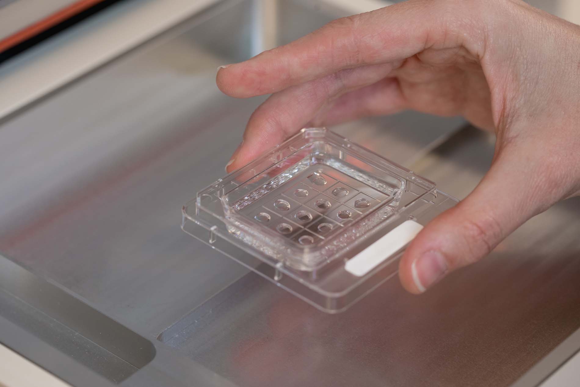
At Equipo Juana Crespo, a sequential approach was devised: first, two cycles of ovarian stimulation with high-precision aspiration targeting the single healthy ovarian pole, avoiding injury, resulting in 9 good-quality embryos. This was followed by a laparoscopic conservative cystectomy, preserving the healthy ovarian parenchyma. Three months later, the patient continued to have regular cycles and preserved ovarian function.
Medical history
The patient had never been pregnant and had not previously undergone any assisted reproduction treatments.
At the age of 18, she underwent emergency laparotomy for a ruptured ovarian cyst with haemoperitoneum (bleeding into the abdominal cavity). A complex cyst was identified in the contralateral ovary and an oophorectomy (complete removal of the ovary) was performed. Pathological examination confirmed a benign dermoid cyst.
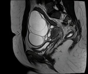
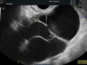
At the age of 28, during a follow-up ultrasound scan, a complex 11 cm mass was detected in the remaining ovary. The magnetic resonance imaging (MRI) was suggestive of a borderline tumour, a neoplasm with low malignant potential for which the standard treatment is surgical. At her original centre, an adnexectomy with staging surgery was proposed, which would have resulted in the loss of her only source of oocytes. The patient came to our clinic to explore alternatives that would preserve her healthy ovarian tissue.
Diagnosis
- Single ovary with an 11 cm complex adnexal mass, suggestive of a borderline tumour.
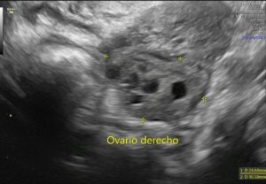
- Healthy ovarian parenchyma confined to the upper pole, with antral follicles (early-stage follicles, indicative of ovarian reserve).
- Right hydrosalpinx (fallopian tube obstructed and dilated by fluid), a factor associated with lower implantation rates.
- Primary infertility for one year and a desire for fertility preservation.
Treatment plan: two-phase fertility preservation
A sequential strategy was proposed: oocyte and embryo preservation first, followed by ovarian-sparing surgery.
Phase 1: precision ovarian aspiration
The main technical difficulty was accessing the healthy parenchyma without aspirating the lesion. As this was a fluid-filled cystic tumour, its components could be mistaken for follicles on ultrasound, and accidental aspiration could compromise the oncological prognosis.
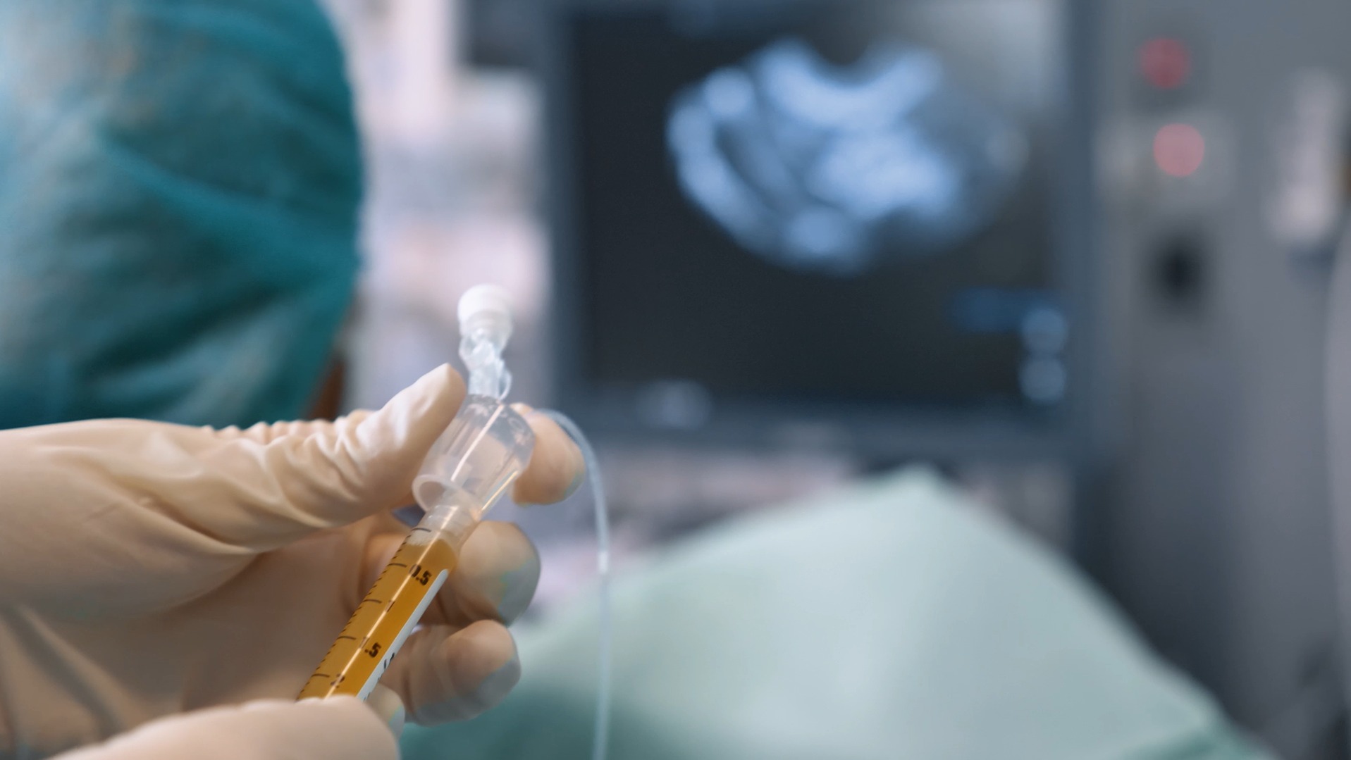
A detailed ultrasound scan enabled us to define the access route to the superior pole of the ovary—the only area containing antral follicles—and to plan each aspiration individually.
Controlled ovarian stimulation
Two cycles of controlled ovarian stimulation were carried out, both without incident:Stage 2: conservative laparoscopic cystectomy
- First cycle: oocyte vitrification for fertility preservation.
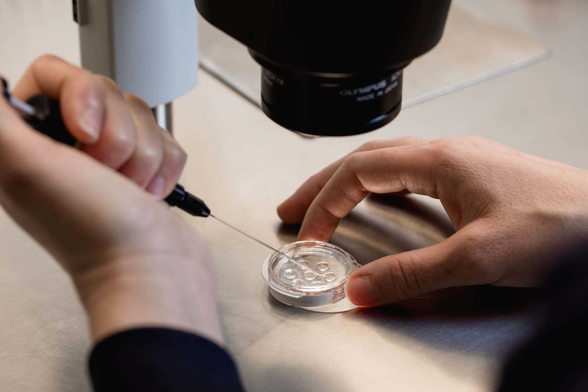
- Second cycle: in vitro fertilisation, with 9 good-quality embryos vitrified.
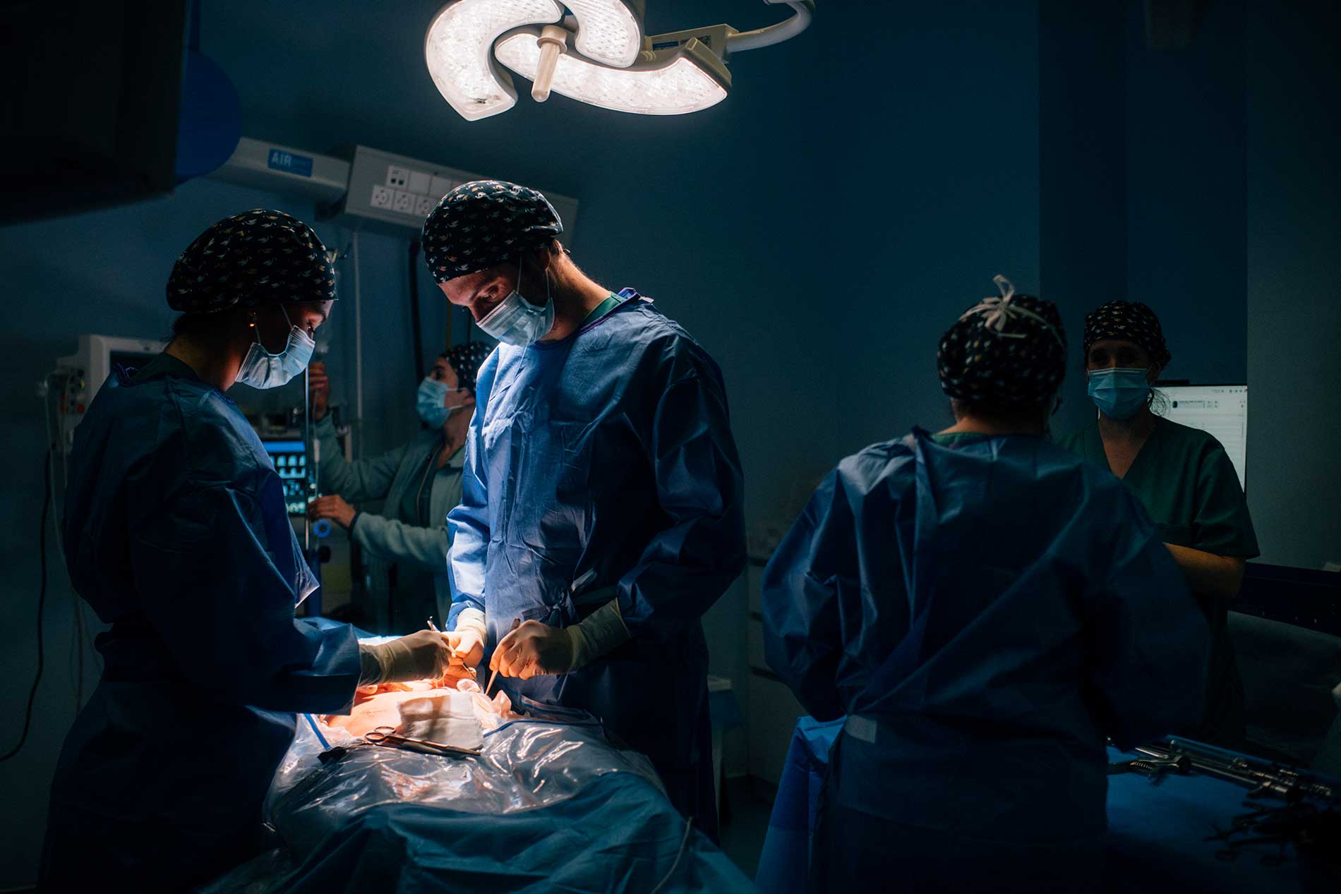
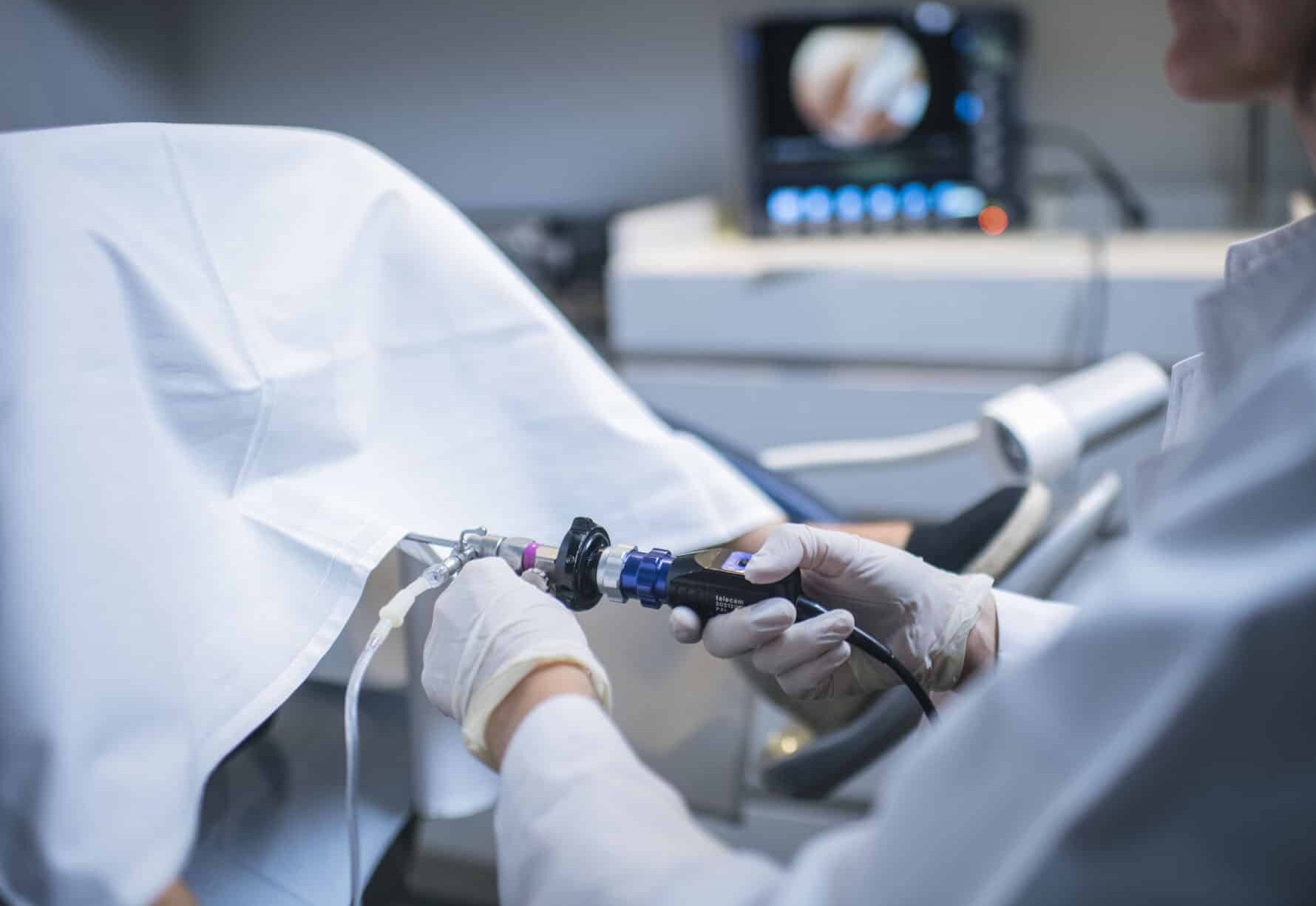
With the embryos already preserved, a laparoscopy was performed with complete resection of the cyst without rupture and preservation of the healthy ovarian parenchyma. This was followed by a bilateral salpingectomy (removal of both fallopian tubes).
The ovary receives blood via two routes: one directly from the ovarian artery and the other from the uterus. To remove the cyst safely, it was necessary to occlude the first of these. The second artery maintains blood supply to the ovary and allows it to continue functioning normally, that is, to produce hormones and mature eggs. As the blood supply is somewhat reduced, follow-up with ultrasound scans and hormone tests was scheduled to check that the ovary retains its function.
Pathological anatomy
The examination ruled out the initial suspicion: the lesion was a mature cystic teratoma, which is benign in nature. Focal chronic salpingitis with paramesonephric cysts was observed in both fallopian tubes, with no oncological significance.
Results
Three months after the operation, the patient had regular menstrual cycles, indicating preserved ovarian function.
The anti-Müllerian hormone (AMH), a marker of ovarian reserve, fell from 2.66 ng/ml before the operation to 1.24 ng/ml afterwards. This decrease is to be expected following the excision of a large lesion and the ligation of one of the ovarian blood supply pathways, and the value obtained suggests a good reproductive prognosis.
With 9 vitrified embryos of good quality and one functional ovary, the couple have decided to postpone the first embryo transfer.
Conclusion
This case demonstrates that, even when cancer is suspected in a single ovary, it is possible to preserve fertility through highly specialised reproductive and surgical planning.
The key was the sequence of treatment: a precision ovarian puncture to retrieve oocytes and embryos without compromising the lesion, followed by conservative surgery that allowed for its complete excision whilst preserving the healthy parenchyma. Thus, the patient retained her ovarian function and the possibility of pregnancy using her own oocytes.


