









Fallo Repetido de Implantación (FRI)
“A successful twin pregnancy using the patient’s own oocytes following repeated implantation failures and a previous egg donation”
Abstract
- Patient Profile: A 25-year-old woman with a history of infertility secondary to tubal/pelvic factors and recurrent implantation failure.
- Medical History: 2 ectopic pregnancies, 1 biochemical pregnancy, 1 early miscarriage, 3 unsuccessful cycles of ovarian stimulation and multiple previous failed cycles of egg donation.
- Diagnostic and Therapeutic Strategy:
1. New course of ovarian stimulation with pre-implantation genetic testing for aneuploidy (PGT-A).
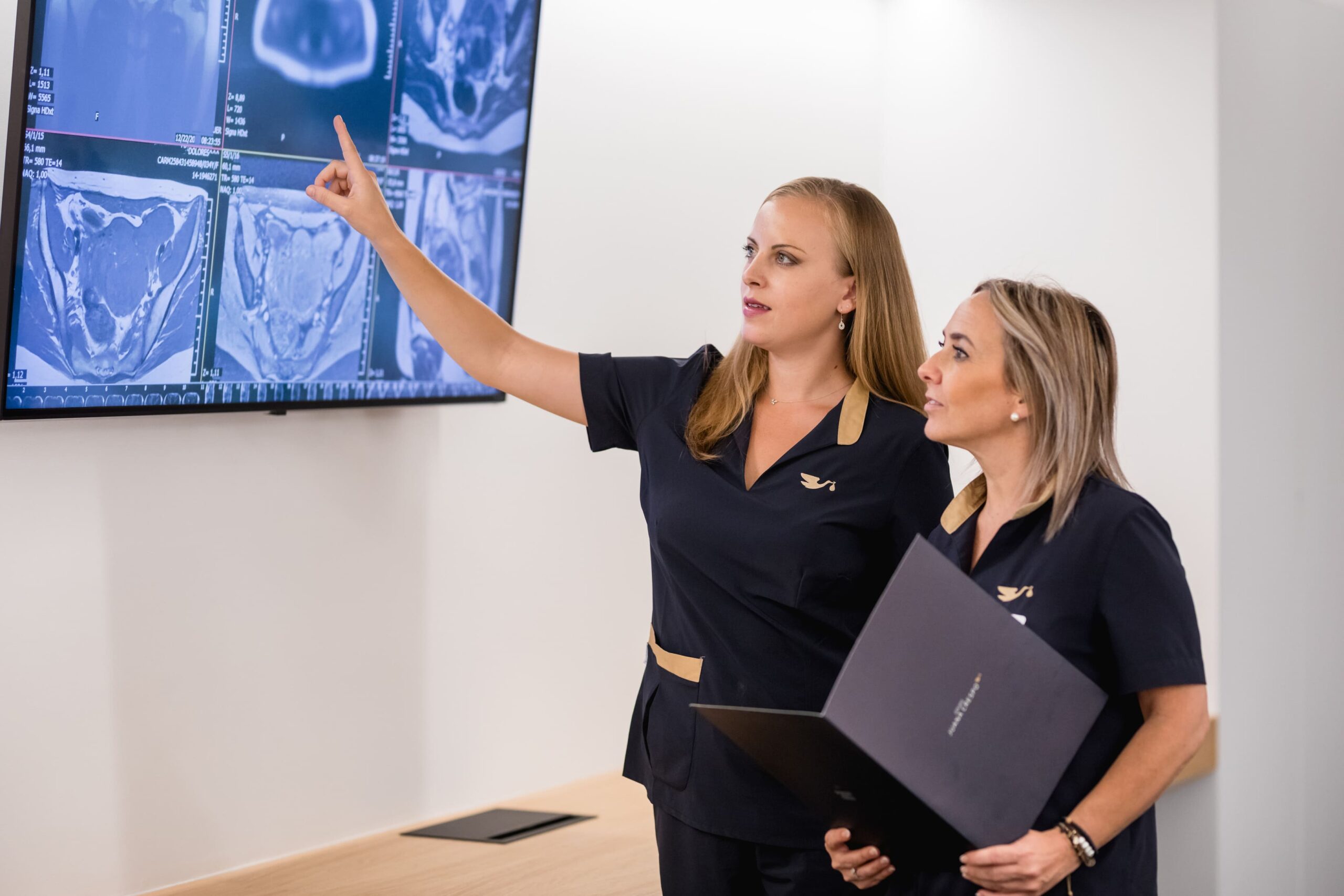
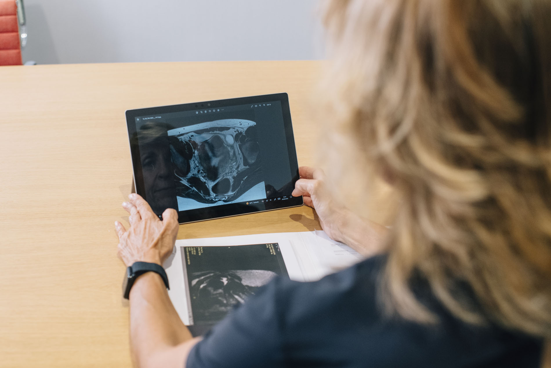
2. Complementary pelvic magnetic resonance imaging (MRI) and hysterosalpingography (HSG).
3. Reconstructive surgical hysteroscopy and endometrial revitalisation with platelet-rich plasma (PRP).
- Obstetric Outcome: Embryo transfer of two euploid embryos performed on 07/07/2026. Confirmation of a progressing dichorionic-biamniotic twin pregnancy (initial β-hCG: 234.94 mIU/ml at 10 days post-transfer).
Reproductive history and medical history
A 25-year-old patient referred to our specialist unit following a long history of unexplained infertility and multiple failures in assisted reproductive technology (ART) treatments.
Previous obstetric history:
- Pregnancy losses: Two previous ectopic pregnancies, one biochemical pregnancy and one early first-trimester pregnancy loss.
- Previous Assisted Reproductive Technology (ART) treatments:
– Three previous cycles of ovarian stimulation for In Vitro Fertilisation (IVF) using her own oocytes, without achieving a viable pregnancy.
– Multiple cycles of embryo transfer using donated oocytes, with recurrent implantation failure.
The medical history suggested not only a possible previous oocyte or embryo quality issue, but also an underlying, undiagnosed uterine or endometrial factor that was preventing successful implantation, regardless of the embryo’s genetic origin.
Multidisciplinary Treatment Strategy
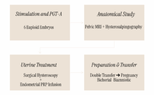
Step 1: Personalised stimulation and genetic testing of the embryos
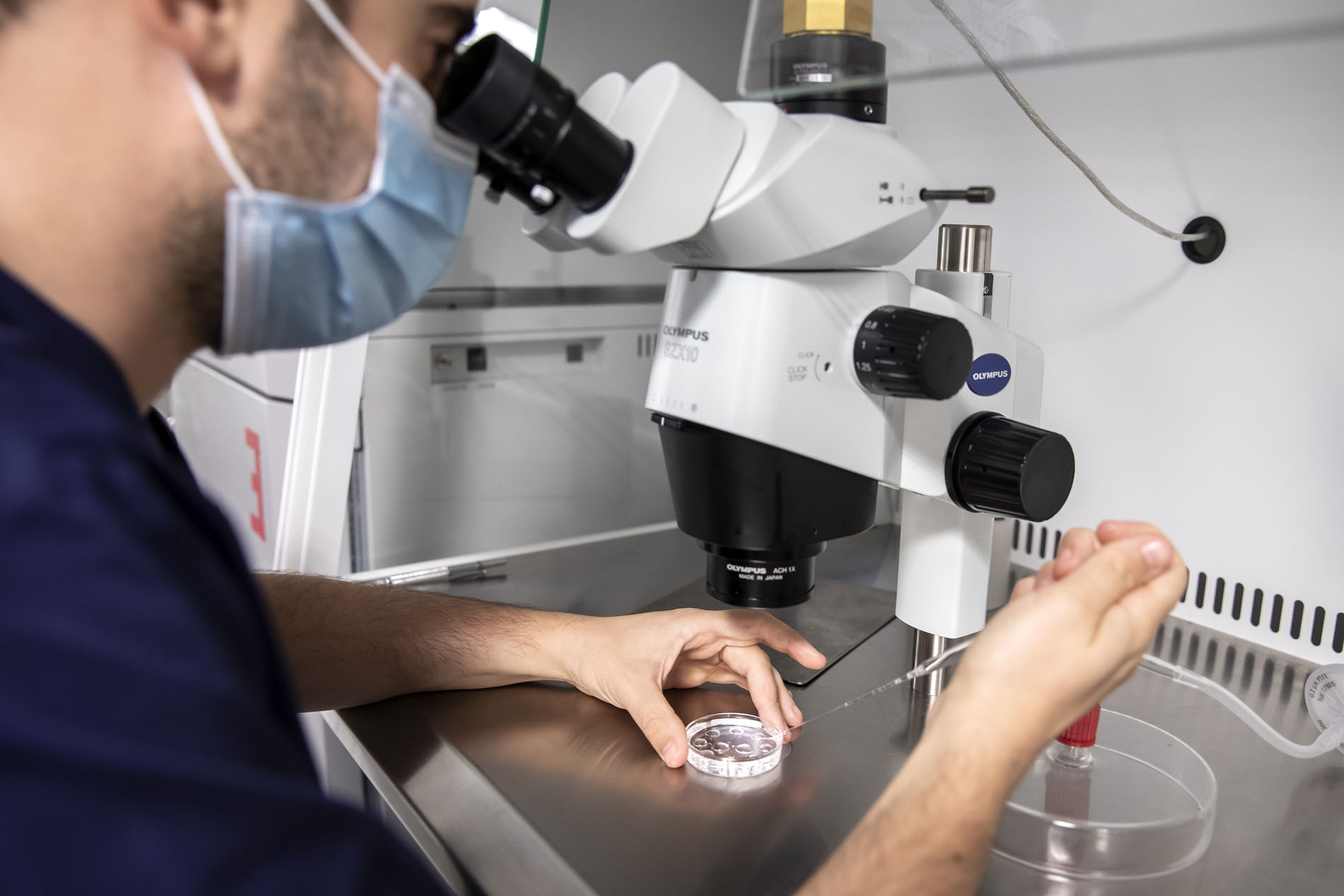
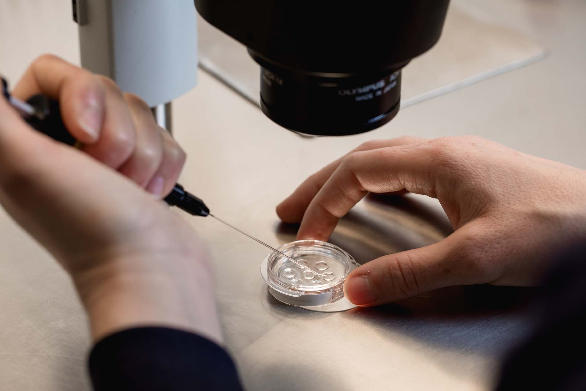
In February 2026, it was decided to carry out a new individualised controlled ovarian stimulation (COS) cycle with the aim of obtaining blastocysts and assessing their chromosomal ploidy via PGT-A (Preimplantation Genetic Testing for Aneuploidy).
- Outcome of the stimulation protocol: Excellent follicular response with the retrieval of 31 oocytes.
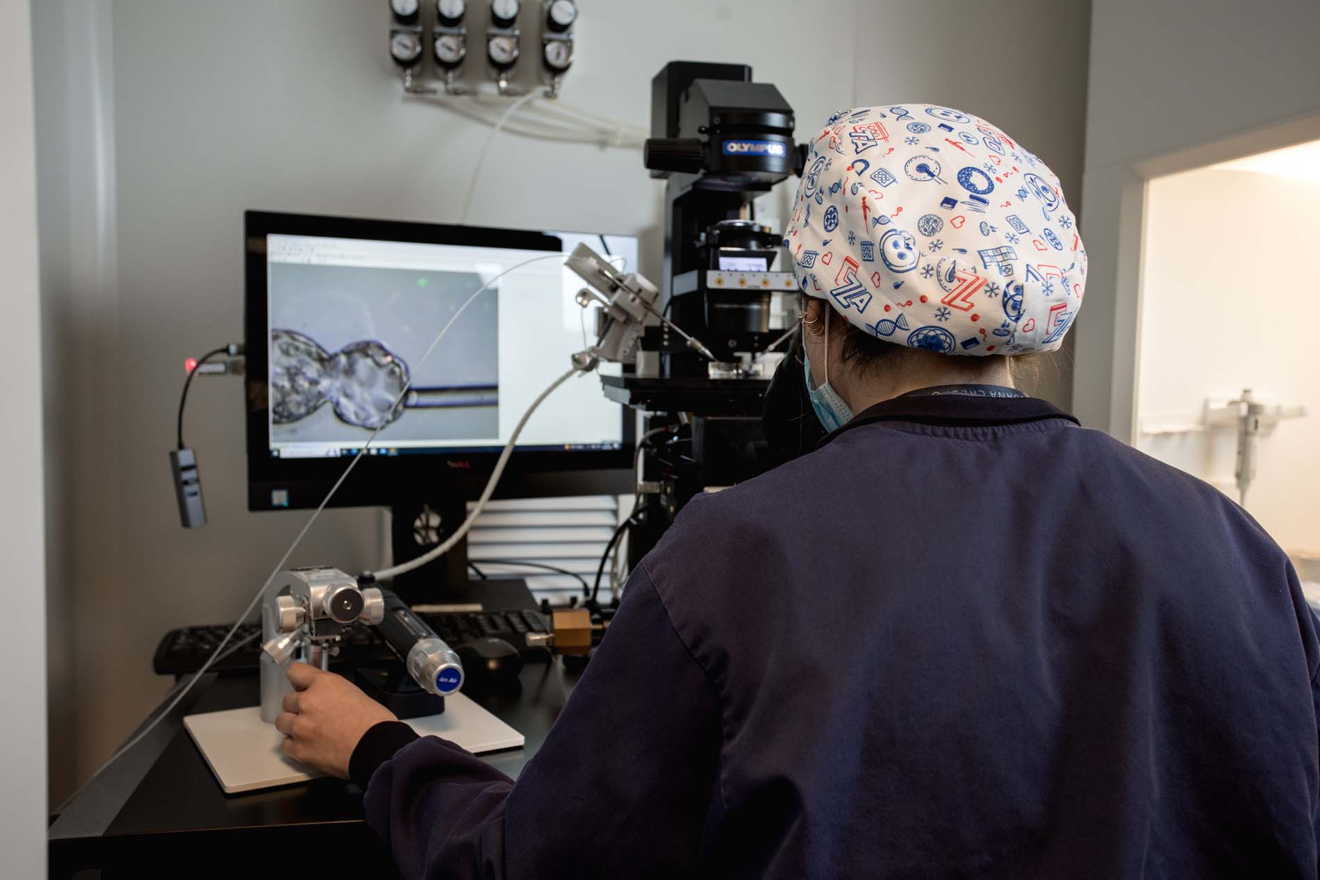
- Embryo development in extended culture: Nine blastocysts of sufficient quality for trophoblast-dermal biopsy were obtained.
- PGT-A results: Six chromosomally normal (euploid) embryos were identified.
This finding confirmed that the patient retained an adequate ovarian reserve and optimal intrinsic reproductive potential, shifting the primary clinical suspicion towards the recipient (uterine/endometrial factor).
Step 2: Endometrial/Uterine Mapping and Anatomical Diagnosis
The morphological assessment of the pelvis was completed using:
Pelvic Magnetic Resonance Imaging (MRI): To rule out deep adenomyosis, Müllerian malformations or subserosal/intramural fibroids.
Hysterosalpingography (HSG): To assess the architecture of the lower uterine segment and the sequelae of previous ectopic pregnancies.
Step 3: Surgical Correction and Endometrial Modulation
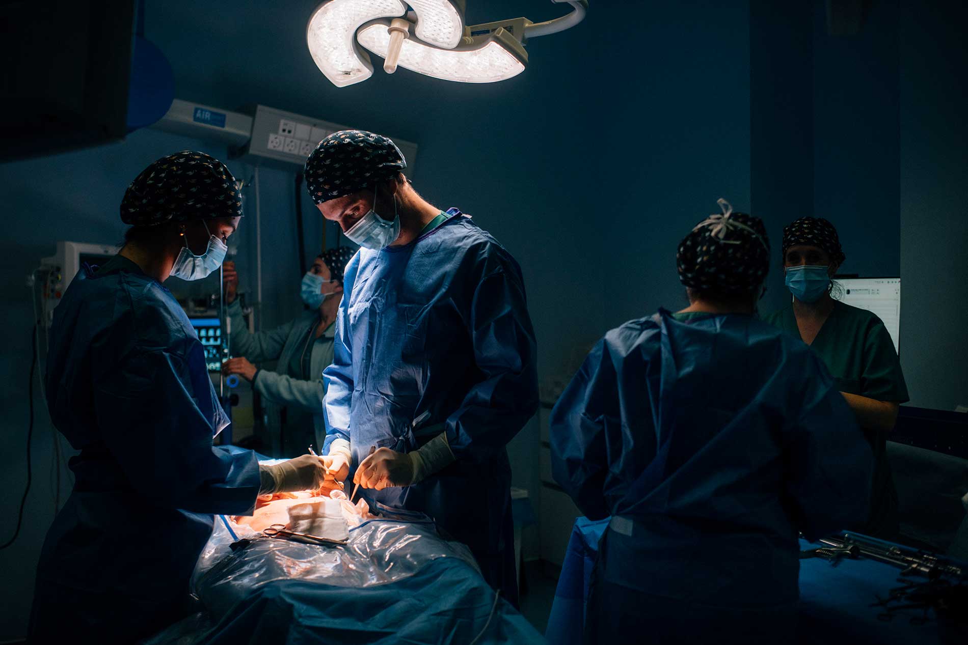
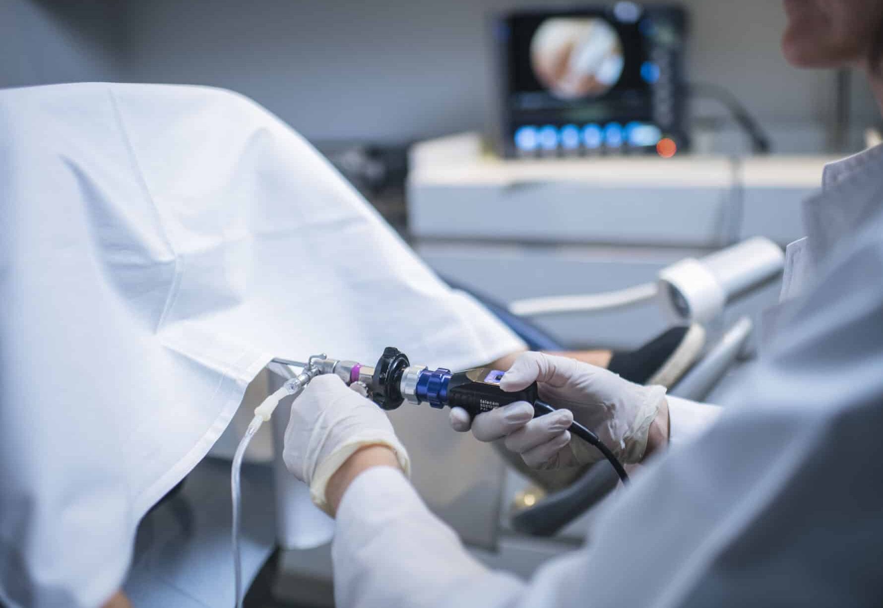
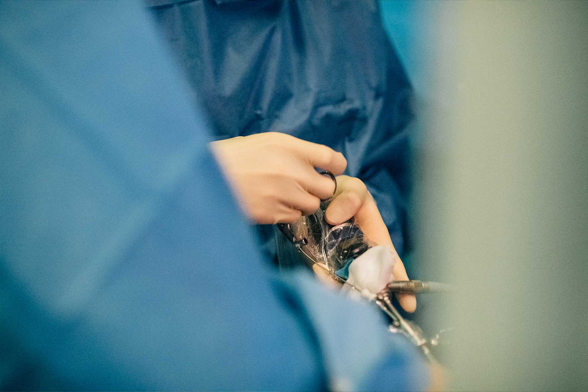
- Surgical Hysteroscopy: Resection and regularisation of the uterine cavity were performed to remove anatomical defects or adhesions (focal Asherman’s syndrome).
- Endometrial Regenerative Medicine (PRP): Intrauterine instillation of platelet-rich plasma (autologous PRP) was administered to restore endometrial thickness, improve subendometrial vascularisation and optimise the implantation window.
Step 4: Endometrial Preparation and Embryo Transfer
Following histological recovery of the uterine cavity, endometrial preparation was initiated using oestrogen and micronised progesterone.
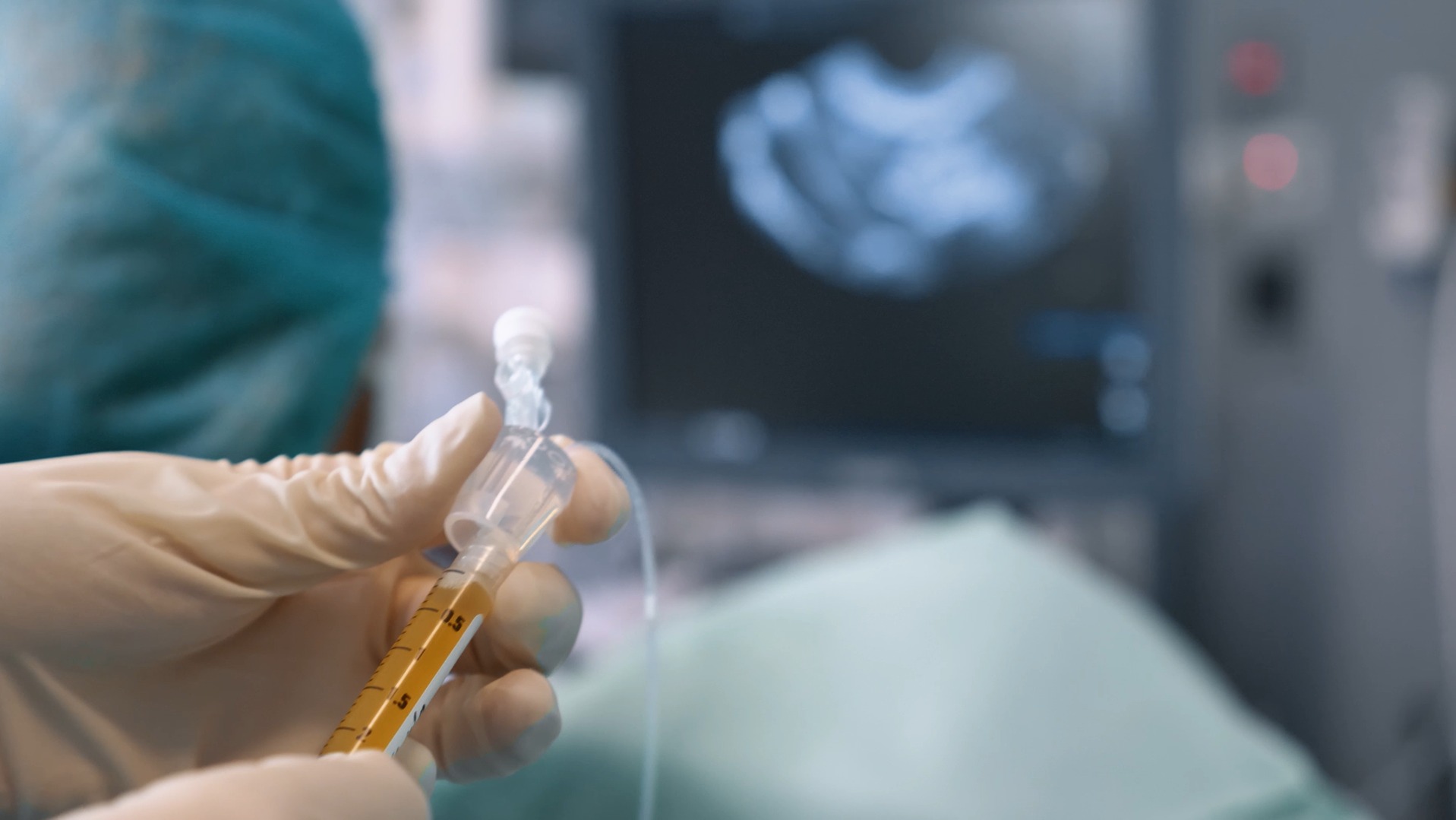
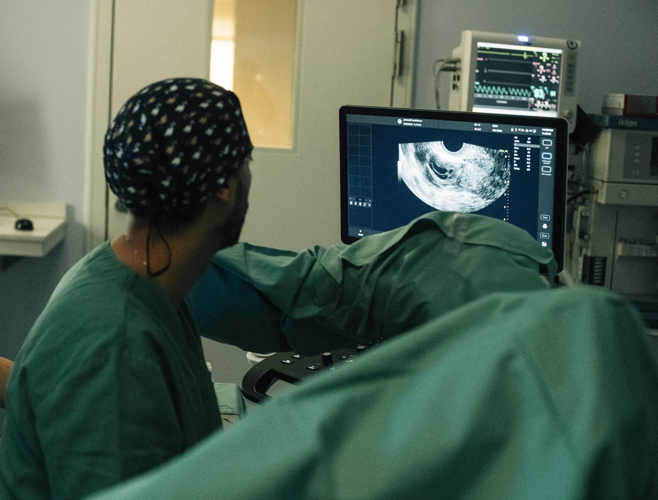
- Transfer Strategy: Two euploid blastocysts were transferred under direct transabdominal ultrasound guidance.
Results and Clinical Course
- Confirmatory Blood Test (β-hCG): On 17/07/2026 (10 days post-transfer), the quantitative β-hCG blood test yielded a positive result of 234.94 mIU/ml.
- Ultrasound Follow-up: The week 6–7 ultrasound scan confirmed the presence of two gestational sacs with yolk sacs and a detectable heartbeat (LCF+), leading to a diagnosis of a developing dichorionic-biamniotic twin pregnancy.
Discussion and Clinical Conclusions
This case demonstrates the importance of comprehensive diagnostic re-evaluation in young patients who have been prematurely labelled as having irreversible reproductive failure.
1. Age vs. Oocyte Quality: Despite previous failed IVF cycles and the use of egg donation, the patient’s biological age (25 years) ensured a high prognosis for embryonic euploidy following optimisation of the stimulation protocol.
2. Relevance of the Uterine Factor in IRF: Implantation failure is not always embryonic in origin; surgical optimisation of the uterine cavity combined with regenerative therapies (PRP) is crucial for restoring endometrial receptivity.
3. Indications for PGT-A: The selection of euploid embryos allows the implantation rate per transfer to be maximised and drastically reduces the time taken to achieve pregnancy.


